Identify the required structures in a published skin image
Inspect an unchanged cutaway illustration and distinguish gland, nerve and fat landmarks.
Section 14.4 · Homeostasis
Exam scope and assessment notes
Extended only (Supplement) · Biology 0970, 2026–2028. Use the stated specimen features and evidence. Diagrams are schematic. Written practice is self-checked; human subject-teacher review, supervised practical competence and cumulative assessment remain pending. Cambridge states that IGCSE 0970 has the same subject content as IGCSE 0610, with 9–1 grading. This StudyForA pathway maps the 2026–2028 syllabus only. This 9–1 graded qualification is available only in a limited number of administrative zones. Confirm that your centre can enter you for this code and exam series with Cambridge International. Cambridge has published a separate syllabus for 2029. StudyForA has not reconciled that version yet; this page supports exams in 2026, 2027 and 2028 only.
Before you begin
- Recall hormone production and blood transport, respiration, liver storage and electrical impulses along neurones.
Work on paper. Try each question before revealing help, and explain why your method works.
Quick readiness check
Which source label corresponds to hair erector muscle?
What you will learn
- Follow the source image’s labelled leaders
- Identify all seven required structures in the cutaway
Identify the required structures in a published skin image
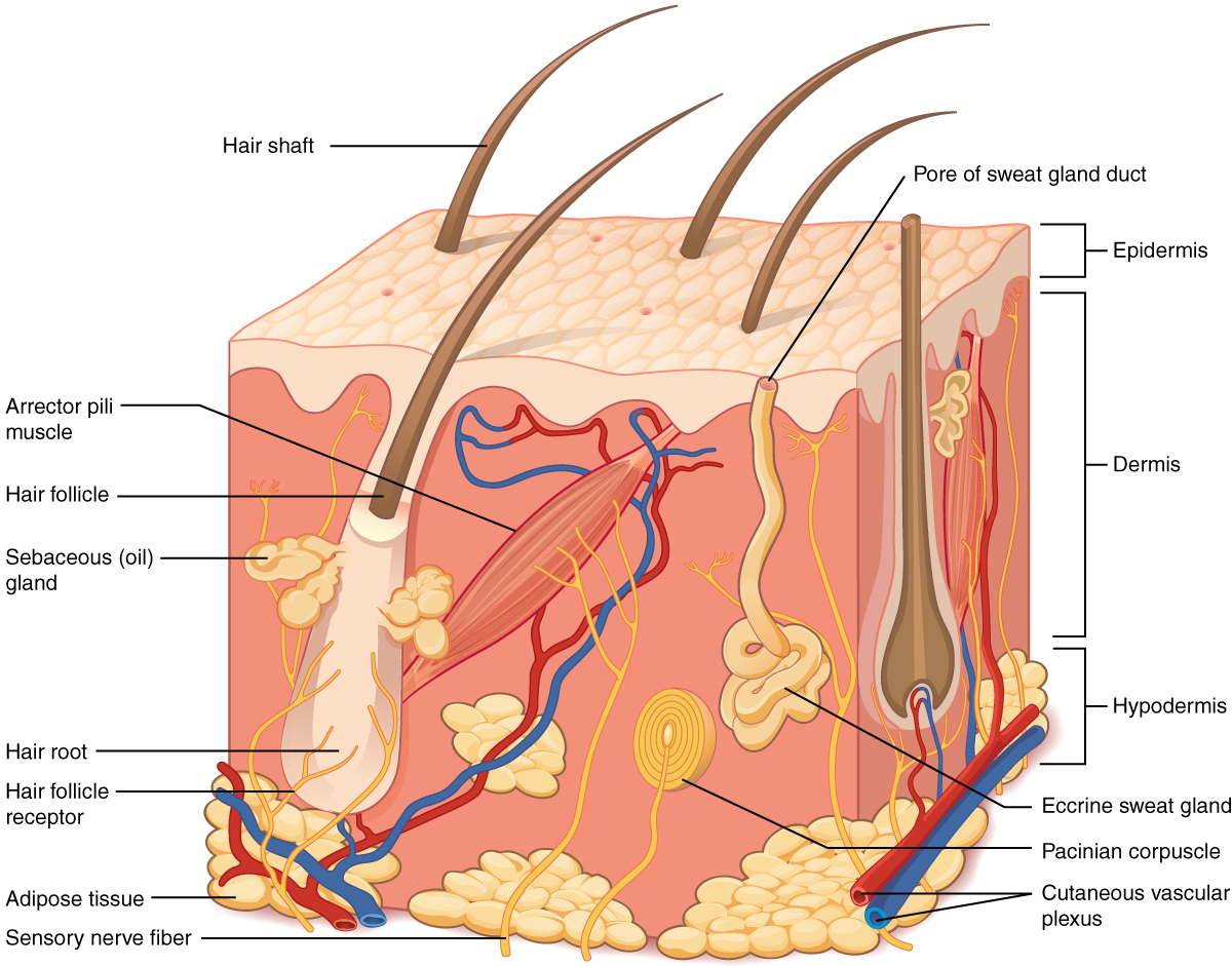
Unchanged OpenStax 2013 hairy-skin cutaway: all seven syllabus structures have labelled landmarks. Illustration, not histology; receptor and sensory process are separate.
Enlarge image for inspection
Inspect the original at its native pixel size. Pan horizontally to follow adjacent labels and leader endpoints.
Check the identification key
Hair: projecting hair shaft. Hair erector muscle: oblique arrector pili band attached to follicle. Sweat gland: right-hand eccrine coil with surface duct, not sebaceous lobules. Receptor: onion-like Pacinian corpuscle or hair follicle receptor. Sensory neurone: separate labelled sensory nerve fiber process; whole cell body omitted. Vessels: lower red/blue cutaneous vascular plexus. Fat: yellow subcutaneous adipose lobules in hypodermis. Colour alone is insufficient; the source does not specifically label thermoreceptors.
Follow the source image’s labelled leaders
The credited OpenStax 2013 illustration is an unchanged published cutaway of hairy skin. Hair shaft projects above the surface. Arrector pili muscle is the oblique red-pink band attached to the follicle, corresponding to the syllabus hair erector muscle. Eccrine sweat gland is the pale coiled tube on the right, with a duct towards the surface sweat pore.
A Pacinian corpuscle is the onion-like sensory receptor low in the image. A hair follicle receptor is also labelled around the root. The sensory nerve fiber label points to a yellow process, representing part of a sensory neurone. Receptors and nerve processes remain distinct; a complete neurone cell body and its full central nervous system route are not shown.
Identify all seven required structures in the cutaway
The cutaneous vascular plexus label identifies the lower red and blue vascular network. Adipose tissue identifies yellow lobules in the hypodermis, the subcutaneous region below the dermis. This published image supplies all seven required identification categories. The hypodermis is associated subcutaneous tissue, rather than being one of the epidermis and dermis main skin layers.
Colour alone is an unreliable key because sweat coils, sensory processes and fat can all look yellowish in the source. Follow labels, shapes, continuity and position together. Distinguish the coiled sweat gland from the sebaceous lobules beside the hair. The source is an anatomical illustration, not histology, and its labelled touch and pressure receptors are not specifically labelled temperature receptors.
Pause and explain
Which source description distinguishes gland and fat?
Worked example
Identify the source landmarks for receptor, sensory neurone and fat.
Show the worked solution
- Pacinian corpuscle is a receptor; sensory nerve fiber is the separate yellow process.
- Adipose tissue is the lobulated subcutaneous fat.
Answer Receptor: Pacinian corpuscle (also hair follicle receptor). Sensory neurone: labelled sensory nerve fiber process. Fatty tissue: adipose lobules in the hypodermis.
Try a topic test
Choose Easy, Normal or Difficult. Save your answers and open worked review after finishing.
Test this topic →From guided practice to a new situation
Write your answer and reasoning first. Use a hint only if you are stuck. The model solution is for self-checking; your written work is not automatically marked.
Name source landmarks for all seven syllabus categories.
Give me a hint
Use the actual printed labels and endpoints.
Compare my reasoning
- Hair shaft; arrector pili muscle; eccrine sweat gland.
- Pacinian corpuscle; sensory nerve fiber; cutaneous vascular plexus; adipose tissue.
Hair: hair shaft. Muscle: arrector pili. Sweat gland: eccrine coil. Receptor: Pacinian corpuscle or hair follicle receptor. Sensory neurone: sensory nerve fiber process. Vessels: cutaneous vascular plexus. Fat: adipose tissue.
Look for these in your work
- Identify all seven categories
- Use source landmarks rather than colour alone
Distinguish the right-hand coiled gland from lower yellow fat and the yellow nerve process.
Give me a hint
Compare shape and connections.
Compare my reasoning
- The coil has a duct to a surface pore.
- Fat is lobulated; the nerve is an elongated branching process.
Sweat gland: coil with a surface duct. Fat: adipose lobules. Sensory nerve: labelled elongated branching process. Shared colour does not make them the same structure.
Look for these in your work
- Use gland continuity
- Distinguish three shapes
Evaluate: this cutaway is a micrograph showing a whole temperature-sensing neurone.
Give me a hint
Check source type and what the labels show.
Compare my reasoning
- It is an anatomical illustration, not a micrograph.
- It shows a sensory process and touch/pressure receptors, not a whole labelled thermoreceptor neurone.
It is a published anatomical illustration. Only a sensory nerve process is shown, and the source does not specifically label the illustrated receptors as temperature receptors.
Look for these in your work
- Identify source type
- Limit the sensory identification claim
Common mistakes
- Calling every yellow structure a sensory neurone.
- Identifying sebaceous lobules as the coiled sweat gland.
What can you explain now?
Which source description distinguishes gland and fat?
Compare with the explanation
Sweat gland is a coil with a duct; adipose tissue is lobulated
Shape and continuity support the identification.
After trying it yourself, choose your next review. This is your self-assessment.
Your review choice appears on Today. Sign in to sync it across devices.
Make it stick
Identify the seven source landmarks and distinguish the limits of a cutaway illustration.
If you needed the solution, close it and solve the problem again from a blank page. A correct answer is stronger when you can explain the reason for each step.