Read genuine dicot leaf cross-section images
Identify resolved tissues and use the source key after attempting the unlabelled image.
Section 6.2 · Leaf structure
Exam scope and assessment notes
Core and Extended · Biology 0970, 2026–2028. Use the stated specimen features and evidence. Diagrams are schematic. Written practice is self-checked; human subject-teacher review, supervised practical competence and cumulative assessment remain pending. Cambridge states that IGCSE 0970 has the same subject content as IGCSE 0610, with 9–1 grading. This StudyForA pathway maps the 2026–2028 syllabus only. This 9–1 graded qualification is available only in a limited number of administrative zones. Confirm that your centre can enter you for this code and exam series with Cambridge International. Cambridge has published a separate syllabus for 2029. StudyForA has not reconciled that version yet; this page supports exams in 2026, 2027 and 2028 only.
Before you begin
- Recall chloroplasts, enzymes and the difference between photosynthesis and respiration.
- Use the stated observations, quantities and controls; diagrams are schematic.
Work on paper. Try each question before revealing help, and explain why your method works.
Quick readiness check
Which evidence is strongest for an air-space label here?
What you will learn
- Identify resolved tissues and use the source key after attempting the unlabelled image.
- Use the stated evidence to explain the method and its limits.
Inspect a genuine dicot leaf section
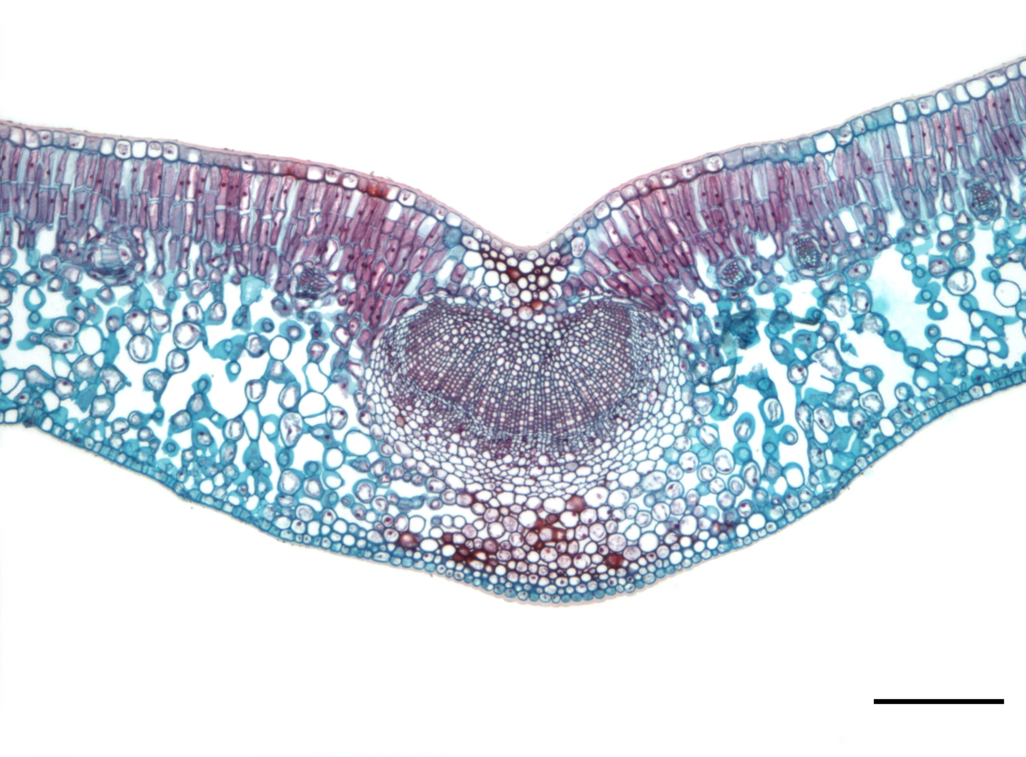
Use the unlabelled microscope image first, then open the source key. A is lower epidermis, B lower palisade, C upper epidermis, D upper palisade, E spongy mesophyll and F a vascular bundle. This specimen has palisade near both surfaces. Individual chloroplasts, cuticle and separate xylem/phloem are not all resolved at this view; lack of resolution does not mean absence.
Check the source image key
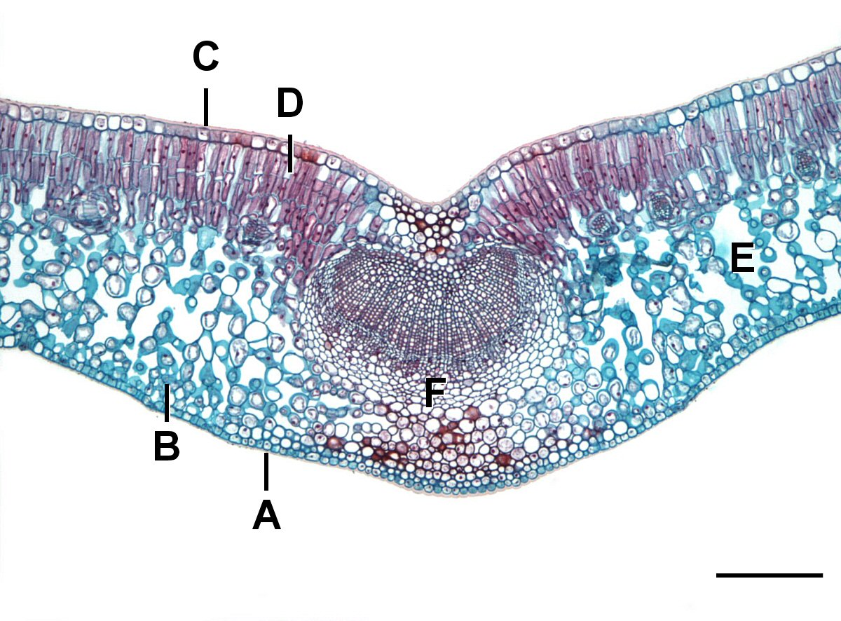
The source has already adjusted colour, brightness, contrast and background. These stain colours are not natural tissue colours. Use the 0.2 mm bar as a ratio; the screen is not a calibrated ruler.
Resolve the cuticle and upper epidermis
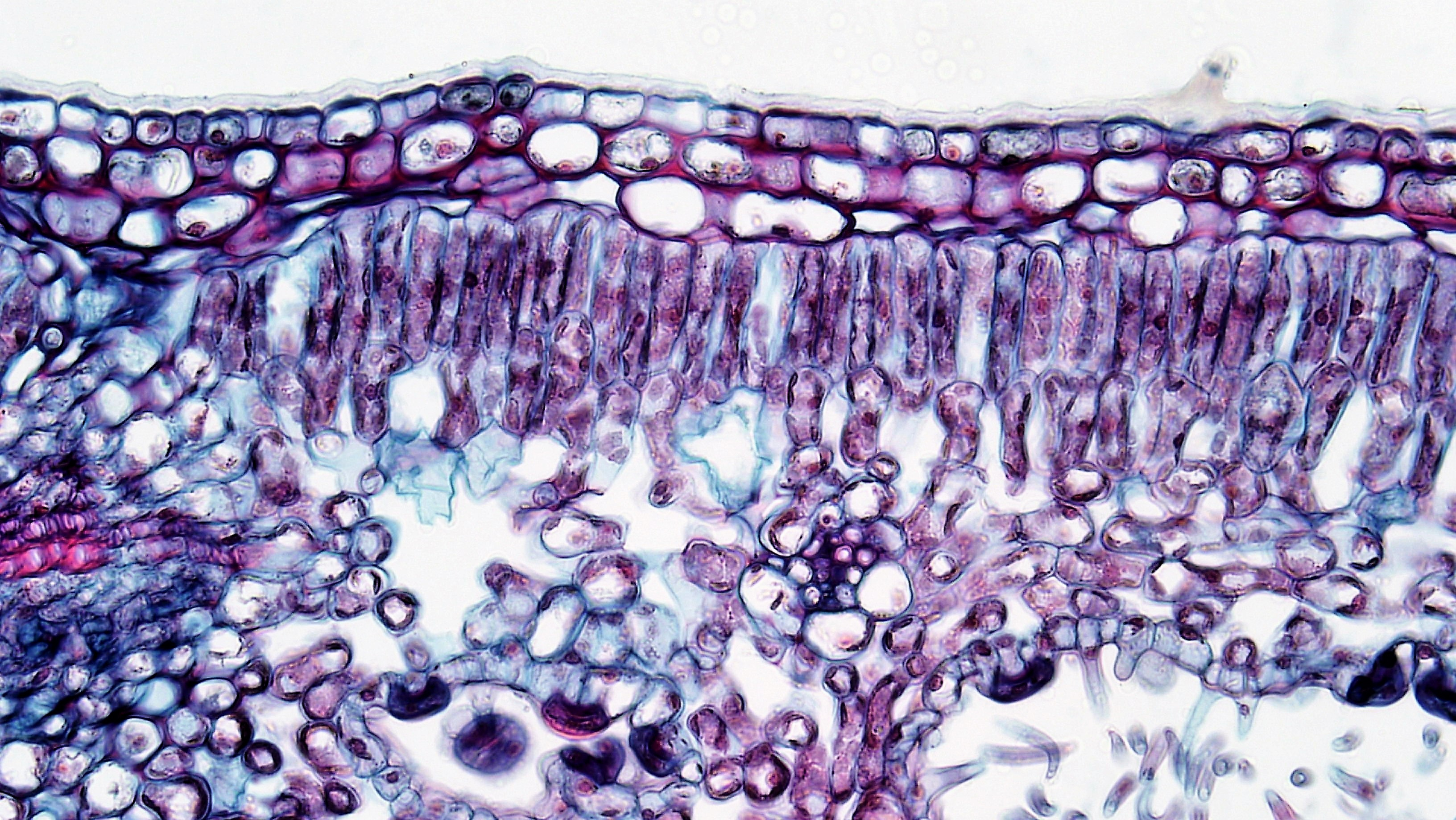
In this Nerium specimen, the outer cuticle follows the top surface. Compact epidermal cells lie immediately beneath it, above elongated palisade cells. The epidermis has several rows in this specimen; do not force it into the single-row class model. Colours are produced by specimen preparation.
Resolve xylem and phloem in a leaf vein
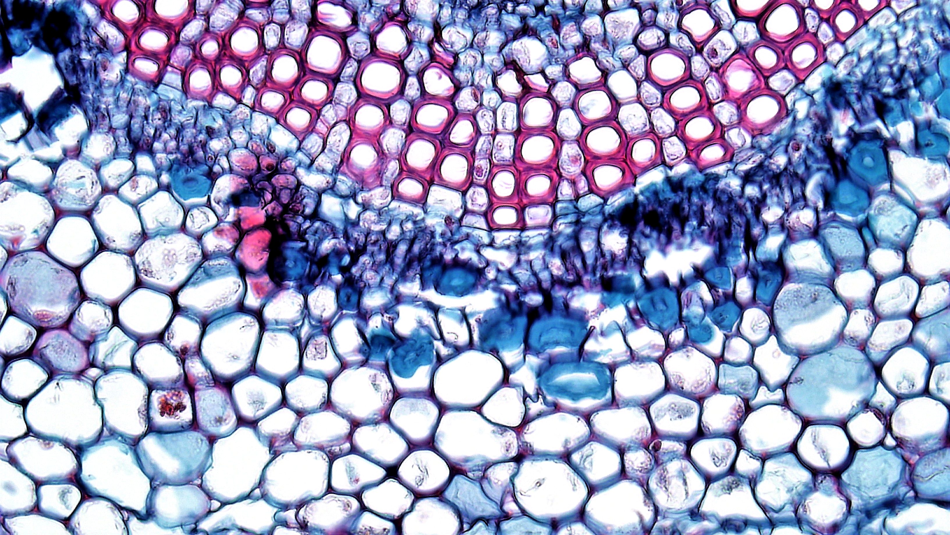
The upper part of this midrib view has large xylem vessel openings. Phloem consists of the smaller cells immediately below that xylem region, with parenchyma around the bundle. Use the documented specimen orientation and structure together. Pink or blue is not a universal tissue key; the source’s nominal ×400 does not calibrate the resized screen.
Start with observable arrangement
The first image is a genuine light-microscope section published by Jon Houseman, with source colour and contrast adjustments retained. Identify the outer cell rows, elongated upper palisade tissue, loosely arranged internal cells and the central cellular vein region. Gaps among spongy cells are air spaces, not missing organelles.
Attempt the unlabelled image before opening the author’s labelled version. In the source key, C identifies upper epidermis, D upper palisade mesophyll, E spongy mesophyll and F the leaf vein. A identifies lower epidermis and B lower palisade tissue in this specimen, which is a reminder that real leaf arrangements can vary.
Use resolution limits honestly
The source scale bar represents 0.2 mm. Resizing the image on screen does not change the represented specimen length; use the bar ratio if a size calculation is requested. Do not use an uncalibrated screen ruler as an absolute specimen-size measurement.
The image supports tissue-level labels but does not clearly resolve every requested feature at every point. Do not claim that all chloroplasts, cuticle thickness or individual xylem and phloem cells are directly identifiable solely by colour here. Use the diagram and a documented detailed image where the structures are resolved. A feature not resolved is not proof that it is absent.
Resolve structures in close-up specimens
The additional Nerium surface image shows a cuticle along the outer top edge, compact upper-epidermal rows below it and elongated palisade cells beneath. This specimen has a multiseriate epidermis: more than one row. A typical diagram is a useful model, but identification must follow the actual specimen.
The separate Nerium midrib image resolves large xylem vessel openings towards the top and smaller phloem cells immediately beneath that xylem region. Parenchyma surrounds the bundle. Combine the documented specimen orientation with structure; stain colour alone is not a general key. No calibrated scale bar is supplied in these close-ups, so do not infer actual dimensions from screen pixels.
Pause and explain
What does failure to resolve an individual chloroplast show?
Worked example
Use the revealed source key to identify C, D, E and F, and distinguish tissue from gaps.
Show the worked solution
- C: upper epidermis; D: upper palisade mesophyll.
- E: spongy mesophyll; F: leaf vein or vascular bundle.
- Air spaces are gaps among the spongy cells, not the whole E tissue.
Answer C upper epidermis; D upper palisade mesophyll; E spongy mesophyll; F vascular bundle.
Try a topic test
Choose Easy, Normal or Difficult. Save your answers and open worked review after finishing.
Test this topic →From guided practice to a new situation
Write your answer and reasoning first. Use a hint only if you are stuck. The model solution is for self-checking; your written work is not automatically marked.
Using the first source key, identify C and D and state one function of D. In the Nerium surface close-up, distinguish the outer cuticle from the compact epidermal rows below it.
Give me a hint
C is a surface layer; D lies beneath it.
Compare my reasoning
- C is upper epidermis.
- D is upper palisade mesophyll.
- Its chloroplast-rich cells support light absorption and photosynthesis.
- Find the outer covering, then the compact cell rows beneath it.
C upper epidermis; D upper palisade mesophyll, supporting photosynthesis. In the close-up, the cuticle is the outer covering; the compact rows immediately below are upper epidermis.
Look for these in your work
- Correct two labels.
- Relevant tissue function.
- Distinguish a surface covering from cells.
Using the first source key, distinguish E, its internal gaps and F. In the Nerium vein close-up, identify the upper large-vessel region and smaller cells immediately beneath it.
Give me a hint
Name tissue, space and vein separately.
Compare my reasoning
- E is spongy mesophyll.
- Gaps among its cells are air spaces.
- F is a leaf vein or vascular bundle.
- Use the documented vein orientation and large vessel openings to locate xylem.
- Locate phloem immediately beneath that xylem region.
E spongy mesophyll; gaps air spaces; F vascular bundle. In the close-up, the upper large-vessel region is xylem; the smaller cells immediately below that region are phloem.
Look for these in your work
- All three identities.
- Avoid calling the whole spongy tissue empty air.
- Locate xylem and phloem from orientation and structure.
The source key labels lower palisade tissue B, but a simple class diagram shows palisade only above. How should the specimen be described?
Give me a hint
A model is not a universal specimen rule.
Compare my reasoning
- Use the image and source key for this specimen.
- Recognise a palisade-like lower region in the supplied key.
- Keep the typical diagram as a simplified arrangement.
Describe the actual keyed lower palisade tissue instead of discarding the evidence to force a simpler model.
Look for these in your work
- Respect specimen evidence.
- Distinguish typical model and variation.
Common mistakes
- Source stains and display colours are not universal tissue codes.
- Do not invent a label for a feature the image does not resolve.
What can you explain now?
What does failure to resolve an individual chloroplast show?
Compare with the explanation
The image does not establish that individual structure clearly.
Resolution limits are not proof of absence.
After trying it yourself, choose your next review. This is your self-assessment.
Your review choice appears on Today. Sign in to sync it across devices.
Make it stick
Identify tissues from arrangement, then check the key and state a resolution limit.
If you needed the solution, close it and solve the problem again from a blank page. A correct answer is stronger when you can explain the reason for each step.