Recognise hepatic vessels in a published liver image
Find the three named routes in a detailed vascular illustration.
Section B9.3 · Blood vessels
Exam scope and assessment notes
Mapped to the named 0973 section using teaching from the corresponding single-science course. A 0973-specific review of every Core and Supplement objective is still in progress. Written practice is self-checked; it does not demonstrate supervised laboratory competence or award an official Cambridge grade. This qualification reports two grades on the 9–1 scale; confirm the entry route for your zone and exam series.
Before you begin
- Recall the original two-inflow liver map.
Work on paper. Try each question before revealing help, and explain why your method works.
Quick readiness check
What kind of image is supplied?
What you will learn
- Use the organ-scale part of the image
- Do not confuse small-scale labels with the required main vessels
Find the three hepatic routes
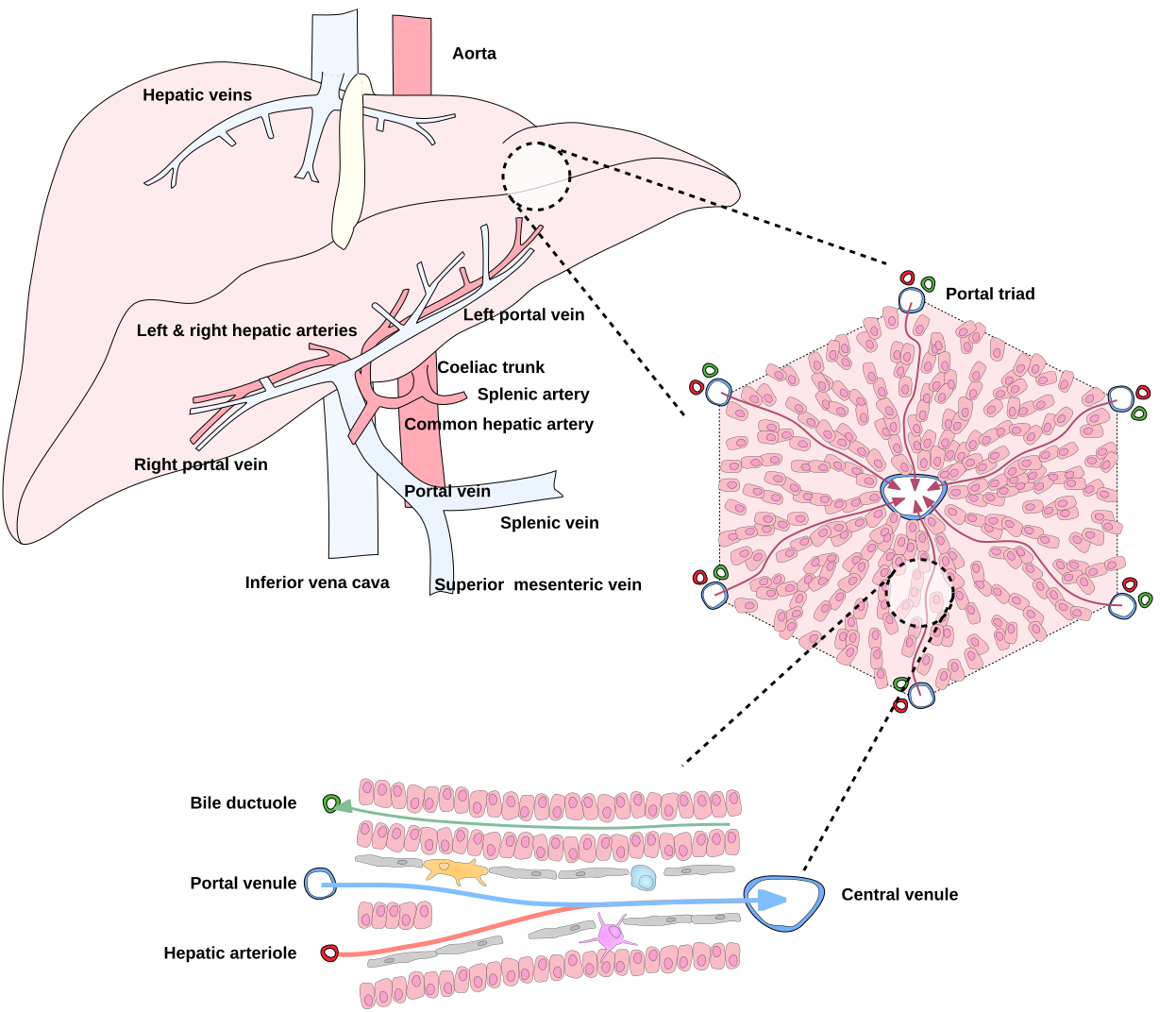
Focus on the left organ-scale liver: hepatic artery, portal vein and hepatic veins. Enlarged lobule labels are supporting context, not extra required main-vessel identifications.
Enlarge image for inspection
Scroll across the enlarged image to inspect labels and cell features. The whole source image is retained. Use the identification key after trying the questions.
Check the identification key
In the left organ-scale image, common hepatic artery branches into left and right hepatic arteries supplying liver. Portal vein enters liver and branches into right and left portal veins: this is hepatic portal inflow from digestive organs. Hepatic veins in the upper liver drain into inferior vena cava. Bile ductuole in the enlarged lower panel is a bile pathway, not a blood vessel. Central venule, portal venule and hepatic arteriole are smaller-scale labels; do not interchange them with the three major vessel names.
Use the organ-scale part of the image
The unchanged illustration by Gwilz includes a liver, its major vessels and enlarged microscopic diagrams. For this identification task, start with the large liver on the left. The common hepatic artery branches into left and right hepatic arteries, supplying the liver. The portal vein enters and branches into right and left portal veins.
Hepatic veins lie in the upper part of the liver and drain into the inferior vena cava. The full-size image keeps these labels legible. Identify each named structure from its label and connection rather than treating every red structure as hepatic artery or every blue structure as hepatic vein.
Do not confuse small-scale labels with the required main vessels
The enlarged panels show portal venules, hepatic arterioles, central venules and a bile ductuole. These help explain the organisation but are not all additional IGCSE main-vessel names to memorise here. A central venule belongs to liver outflow inside a lobule; it is not a label for the entire hepatic portal vein.
The bile duct transports bile and is not a blood vessel. Follow the major vessels in the left organ-scale image to distinguish arterial inflow, portal inflow and hepatic-vein outflow. This is a published anatomical illustration, not a tissue photomicrograph or direct evidence of a learner’s practical dissection competence.
Sources for this lesson
Pause and explain
Which labels identify the arterial inflow in the liver panel?
Worked example
In the left liver image, identify the upper drainage branches and the vessel receiving them.
Show the worked solution
- Read “hepatic veins” in the upper liver region.
- Trace their connection to the inferior vena cava.
Answer Hepatic veins drain the liver into the inferior vena cava.
Try a topic test
Choose Easy, Normal or Difficult. Save your answers and open worked review after finishing.
Test this topic →From guided practice to a new situation
Write your answer and reasoning first. Use a hint only if you are stuck. The model solution is for self-checking; your written work is not automatically marked.
Locate and name the arterial and portal inflows in the left organ-scale image.
Give me a hint
Use common hepatic artery and portal vein labels.
Compare my reasoning
- The arterial inflow is the common hepatic artery and its hepatic branches.
- The portal inflow is the portal vein and its branches.
Hepatic artery supplies arterial blood; hepatic portal vein brings digestive-organ blood into liver.
Look for these in your work
- Identify both image routes
- Distinguish their origins
Locate hepatic veins and state where they drain.
Give me a hint
Look in the upper liver and follow the receiving vessel.
Compare my reasoning
- Hepatic veins leave the liver.
- Their drainage enters the inferior vena cava.
Hepatic veins → inferior vena cava.
Look for these in your work
- Identify the outflow label
- Name its receiving vessel
Why should an enlarged green bile-ductuole symbol not be named hepatic portal vein?
Give me a hint
Compare the label, carried material and vessel connection.
Compare my reasoning
- The ductuole is labelled as a bile pathway.
- Hepatic portal vein is the major digestive-organ blood inflow.
The symbol is a bile ductuole, not a blood vessel; identify hepatic portal vein from the labelled blood inflow to liver.
Look for these in your work
- Use the actual label
- Distinguish bile and blood
Common mistakes
- Labelling a bile ductuole as hepatic artery.
- Using portal vein and hepatic veins as the same name.
What can you explain now?
Which labels identify the arterial inflow in the liver panel?
Compare with the explanation
Common hepatic artery and left/right hepatic arteries
These are arterial branches supplying liver.
After trying it yourself, choose your next review. This is your self-assessment.
Your review choice appears on Today. Sign in to sync it across devices.
Make it stick
Use the full-size image to identify all three major routes without the key.
If you needed the solution, close it and solve the problem again from a blank page. A correct answer is stronger when you can explain the reason for each step.