Distinguish a receptor from a sensory neurone in an image
Use a complementary thick-skin illustration without treating it as a crop of the hairy-skin source.
Section 14.4 · Homeostasis
Extended only (Supplement); Biology 0610, 2026–2028. Use the stated specimen features and evidence. Diagrams are schematic. Written practice is self-checked
Before you begin
- Recall hormone production and blood transport, respiration, liver storage and electrical impulses along neurones.
Work on paper. Try each question before revealing help, and explain why your method works.
Quick readiness check
Which label identifies the blue basal receptor?
What you will learn
- Identify the Merkel receptor and nerve process
- Compare what the two published images include
Distinguish a receptor from a sensory neurone in an image
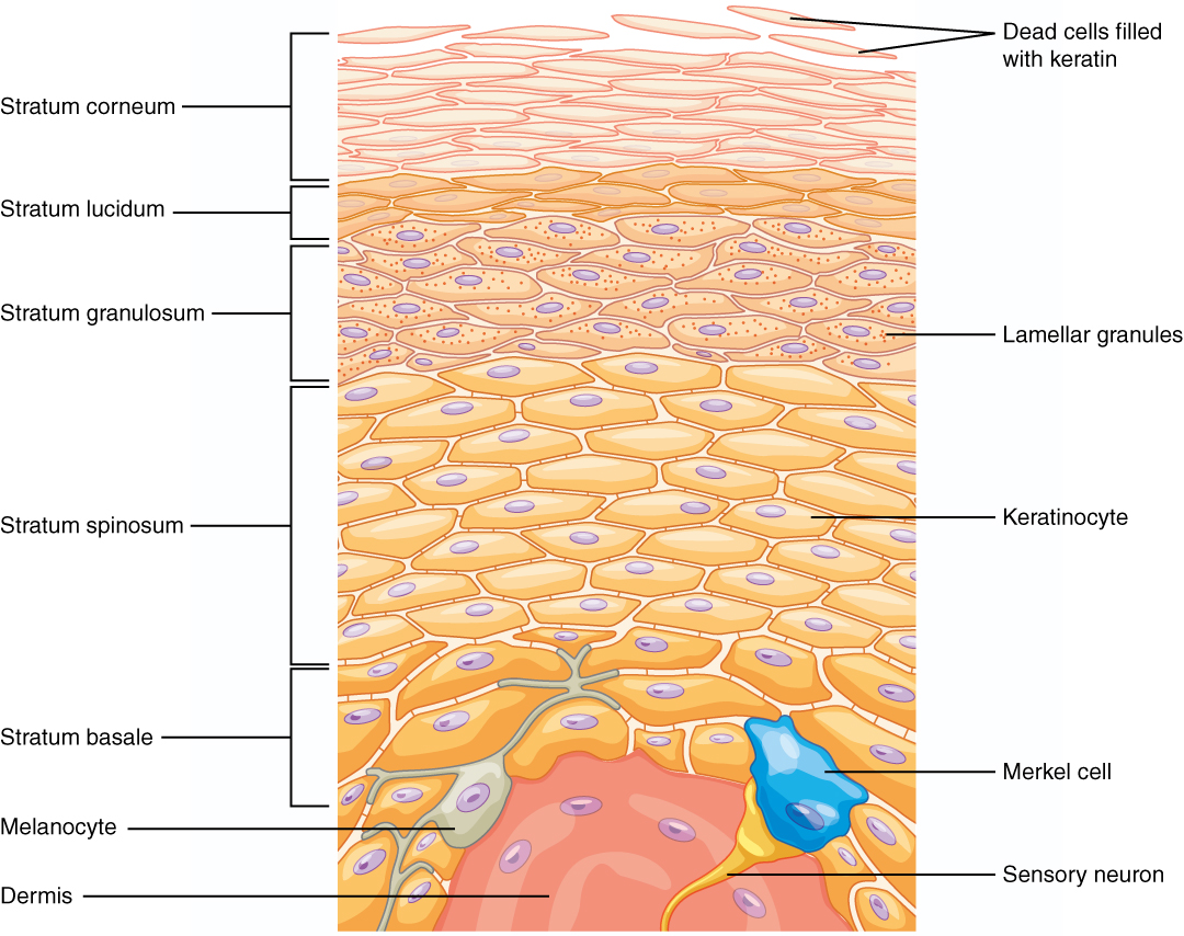
Unchanged OpenStax 2013 thick-skin epidermis: blue Merkel receptor and separate yellow sensory nerve process. Not a crop of the main hairy-skin image; whole neurone omitted.
Enlarge image for inspection
Inspect the original at its native pixel size. Pan horizontally to follow adjacent labels and leader endpoints.
Check the identification key
Merkel cell: blue touch-associated receptor near the epidermal base. Sensory neuron: separate yellow process contacting it; whole cell body and full CNS route absent. Thick-skin epidermis, not a magnified crop of the hairy-skin image. Hair, erector muscle, sweat gland, vessels and fat are absent; use the main cutaway for those five categories. Neither label specifically identifies a temperature receptor.
Identify the Merkel receptor and nerve process
The complementary OpenStax 2013 illustration focuses on epidermal layers of thick skin. A blue Merkel cell is labelled near the base of the epidermis. It is a touch-associated receptor cell, contacting a distinct yellow sensory nerve terminal. The separate sensory neuron leader points to the yellow process below it rather than to the blue cell.
The receptor detects an appropriate stimulus and the sensory neurone transmits electrical impulses towards the central nervous system. The yellow structure drawn in this image is a process of that neurone; its whole cell body and complete route are outside the image. Distinct labels should not be merged into one name simply because the two structures touch.
Compare what the two published images include
This is a complementary image of thick-skin epidermis, not a magnified crop of the hairy-skin cutaway. It includes the stratum lucidum associated with thick skin. It does not supply the other five required categories of hair, hair erector muscle, sweat gland, blood vessels and fatty tissue, so use the main cutaway for those structures.
When comparing sources, report both the useful identification and the limitation. The image supports distinguishing Merkel cell from sensory nerve process, but does not specifically label either as the temperature detector used in the later thermoregulation model. Identifying a touch receptor is valid for the general skin-structure requirement without inventing its stimulus specificity.
Pause and explain
Can this complement alone identify all seven required categories?
Worked example
Identify the two labelled structures and the image’s scope.
Show the worked solution
- Blue basal Merkel cell is the receptor; yellow sensory neuron label reaches the nerve process.
- The illustration is thick-skin epidermis, not a crop supplying all seven structures.
Answer Merkel cell: blue receptor near the epidermal base. Sensory neuron: separate yellow process. This thick-skin illustration complements the main cutaway; it does not show all seven categories.
Try a topic test
Choose Easy, Normal or Difficult. Save your answers and open worked review after finishing.
Test this topic →From guided practice to a new situation
Write your answer and reasoning first. Use a hint only if you are stuck. The model solution is for self-checking; your written work is not automatically marked.
Distinguish the Merkel cell and sensory neuron endpoints.
Give me a hint
Follow both printed leaders independently.
Compare my reasoning
- Merkel cell points to a blue basal receptor cell.
- Sensory neuron points to a yellow nerve process.
Merkel cell: blue basal receptor cell. Sensory neuron: separate yellow nerve process, not the whole cell body.
Look for these in your work
- Identify each endpoint
- Avoid claiming a whole neurone
Name the five required categories for which the main image is still needed.
Give me a hint
Exclude receptor and sensory neurone, which this complement supports.
Compare my reasoning
- Hair, hair erector muscle and sweat gland are needed.
- Blood vessels and fatty tissue are also needed.
Hair, hair erector muscle, sweat gland, blood vessels and fatty tissue.
Look for these in your work
- List all five missing categories
- Use the complementary sources together
Why does identifying a Merkel cell not establish the specific thermoreceptor in temperature control?
Give me a hint
Use the source’s stated touch association.
Compare my reasoning
- Merkel cell is a touch-associated receptor.
- This source does not label a particular temperature receptor.
It identifies a touch-associated receptor, not a specifically labelled temperature receptor. Do not infer stimulus specificity beyond the source.
Look for these in your work
- State the source association
- Limit the thermoreceptor claim
Common mistakes
- Calling the yellow process the blue Merkel cell.
- Treating thick-skin epidermis as a direct zoom of the hairy-skin cutaway.
What can you explain now?
Can this complement alone identify all seven required categories?
Compare with the explanation
No; use the main cutaway for the other five
Its narrower scope is receptor and sensory-process identification.
After trying it yourself, choose your next review. This is your self-assessment.
Your review choice appears on Today. Sign in to sync it across devices.
Make it stick
Identify the separate receptor and sensory process and state why the complementary image cannot replace the main source.
If you needed the solution, close it and solve the problem again from a blank page. A correct answer is stronger when you can explain the reason for each step.