Identify organs in a published digestive-system image
Use pointers and connections before opening the identification key.
Section 7.2 · Digestive system
Core and Extended; Biology 0610, 2026–2028. Use the stated specimen features and evidence. Diagrams are schematic. Written practice is self-checked
Before you begin
- Recall large and small nutrient molecules, cell membranes and enzyme specificity.
- Use the stated diagram keys and evidence; models are not to scale.
Work on paper. Try each question before revealing help, and explain why your method works.
Quick readiness check
Which evidence helps identify an organ in an unfamiliar picture?
What you will learn
- Use pointers and connections before opening the identification key.
- Interpret a published illustration without claiming it measures organ function.
Try the numbered organ pointers
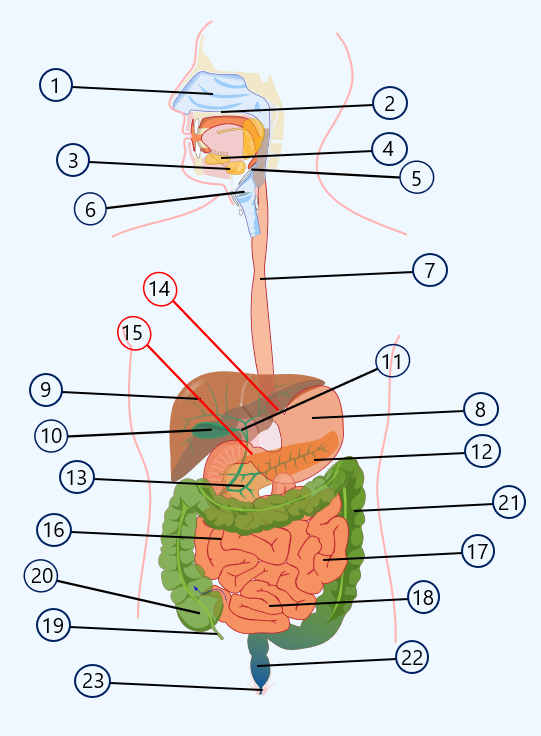
The source is an anatomical illustration, not a photograph or a measurement of organ function. Attempt the named organs before opening the key. Use pointer endpoints and connections; not all numbered structures are required by the syllabus.
Check the identification key
Required structures: mouth region at the upper opening; 3–5 salivary glands; 7 oesophagus; 8 stomach; 9 liver; 10 gallbladder; 12 pancreas; 13 duodenum; 18 ileum; 21 colon; 22 rectum; 23 anus. The unnumbered English-labelled source below verifies the anatomical endpoints. It uses “esophagus” for oesophagus. Other numbered structures are outside the required list.
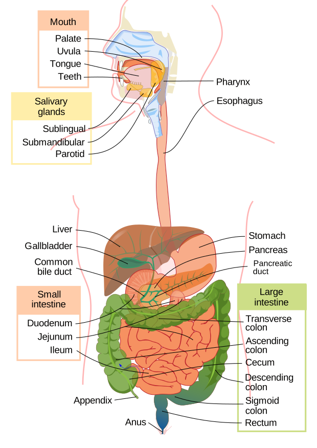
The two published versions differ in labelling and framing. Follow endpoints rather than assuming pixel locations match.
Inspect the source image
The published anatomical illustration uses numbered pointers. Begin with the long tube from the mouth region to the stomach, the liver above the gut coils, the gallbladder beneath the liver, and the pancreas near the duodenum. Do not identify a structure solely by its fill colour.
The image represents anatomy, not a photograph of a living person or a measurement of organ size. Some pointers mark structures outside the required syllabus list. Use the separate key only after attempting the named organs.
Distinguish similar regions
The duodenum is the first small-intestine region around the pancreatic area; the ileum is the final small-intestine region before the large intestine. Colon surrounds much of the small-intestine coils in this front view. Rectum and anus form the final route below them.
Compare the image with the original route model. The original model helps with function and sequence; the source image adds anatomical layout. Neither proves movement direction from a still frame alone.
Pause and explain
Which numbered image organ is part of the food canal?
Worked example
Identify the organ at pointer 8 and distinguish it from the organ at pointer 9.
Show the worked solution
- Pointer 8 enters the stomach sac.
- Pointer 9 enters the liver above it.
- Only the stomach is on the swallowed-food route.
Answer 8: stomach; 9: liver. The stomach is a canal organ, while the liver supplies bile.
Try a topic test
Choose Easy, Normal or Difficult. Save your answers and open worked review after finishing.
Test this topic →From guided practice to a new situation
Write your answer and reasoning first. Use a hint only if you are stuck. The model solution is for self-checking; your written work is not automatically marked.
Identify pointers 7, 8 and 9 in the source image, then check the key.
Give me a hint
Trace the tube and nearby organs.
Compare my reasoning
- 7 is the oesophagus.
- 8 is the stomach.
- 9 is the liver.
7: oesophagus; 8: stomach; 9: liver.
Look for these in your work
- All three.
- Use endpoints.
Identify the small bile-storage sac and the gland that supplies pancreatic enzymes in the source image.
Give me a hint
Look beneath the liver and beside the duodenum.
Compare my reasoning
- 10 identifies gallbladder.
- 12 identifies pancreas.
10: gallbladder; 12: pancreas.
Look for these in your work
- Both organs.
- Separate bile and enzymes.
Use pointers 13, 18, 21, 22 and 23 to trace required intestinal regions in the image.
Give me a hint
Use the source key after your attempt.
Compare my reasoning
- 13: duodenum.
- 18: ileum; 21: colon.
- 22: rectum; 23: anus.
13 duodenum → onward to 18 ileum → 21 colon → 22 rectum → 23 anus.
Look for these in your work
- All named regions.
- Treat the picture as an anatomical illustration.
Common mistakes
- An illustration is not a live physiological measurement.
- Pointer endpoints matter more than colour.
What can you explain now?
Which numbered image organ is part of the food canal?
Compare with the explanation
8, the stomach.
Food enters the stomach.
After trying it yourself, choose your next review. This is your self-assessment.
Your review choice appears on Today. Sign in to sync it across devices.
Make it stick
Identify the canal and associated organs without relying on a fixed colour key.
If you needed the solution, close it and solve the problem again from a blank page. A correct answer is stronger when you can explain the reason for each step.