Identify urinary organs in a published system image
Trace numbered source leaders to kidneys ureters bladder and urethra.
Section 13.1 · Excretion in humans
Core and Extended; Biology 0610, 2026–2028. Use the stated specimen features and evidence. Diagrams are schematic. Written practice is self-checked
Before you begin
- Recall cell metabolism, blood transport, amino acids and the distinction between excretion and egestion.
Work on paper. Try each question before revealing help, and explain why your method works.
Quick readiness check
What is the source image type?
What you will learn
- Use the unchanged source numbers
- Distinguish extra structures from the urine route
Identify urinary organs in a published system image
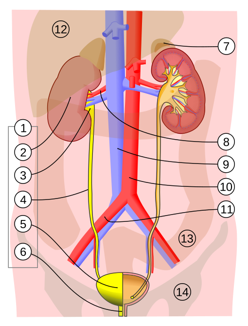
Unchanged published illustration: identify numbers 2, 4, 5 and 6 before opening the key. These correspond to kidney, ureter, bladder and urethra.
Enlarge image for inspection
Scroll across the source to trace each leader or bracket. Try identification before opening the key.
Check the identification key
Required source numbers: 2 kidney; 4 ureter; 5 bladder; 6 urethra. The source’s extra number 3 is renal pelvis and 8 identifies renal blood vessels. Numbers 2, 4, 5 and 6 correspond to A, B, C and D in our separate original diagram; those letters are not added to this unchanged source.
Use the unchanged source numbers
The published urinary-system illustration by Jordi March i Nogué retains its original numbered leaders. Number 2 points into the kidney on the image-left side; 4 points to its descending ureter; 5 ends inside the bladder; and 6 points to the short outlet below it, the urethra. The source numbers are not the A–D letters used in our separate original diagram.
Follow each leader to its endpoint rather than using the position of the number circle. The paired kidneys and ureters are visible, while one kidney and the bladder are partly sectioned to expose internal features. The urine route proceeds from kidney through ureter to bladder and then urethra. The image is a published anatomical illustration, not a specimen photograph.
Distinguish extra structures from the urine route
The source contains extra organs, regions and blood vessels. Number 3 identifies the renal pelvis, so it should not replace 4 as the ureter answer. Red and blue renal blood vessels are a circulation route rather than the yellow urine tubes. Other numbered structures help orientation but are not the four required identification targets in this activity.
The short depicted urethral outlet is simplified and does not teach exact anatomical length or differences between people. Try the four required numbers before opening the separate key, and enlarge the source if a leader is difficult to follow. Keep the source labels intact and avoid inferring a scale measurement, a sex or an individual medical condition from this teaching view.
Pause and explain
Which source number identifies the ureter?
Worked example
Identify source numbers 2, 4, 5 and 6.
Show the worked solution
- Trace 2 into the kidney and 4 to its descending tube.
- Trace 5 into storage and 6 below the bladder.
Answer 2 kidney; 4 ureter; 5 bladder; 6 urethra.
Try a topic test
Choose Easy, Normal or Difficult. Save your answers and open worked review after finishing.
Test this topic →From guided practice to a new situation
Write your answer and reasoning first. Use a hint only if you are stuck. The model solution is for self-checking; your written work is not automatically marked.
Identify all four target source numbers from their leader endpoints.
Give me a hint
Keep the original numerical key.
Compare my reasoning
- 2 indicates kidney and 4 ureter.
- 5 indicates bladder and 6 urethra.
2 kidney; 4 ureter; 5 bladder; 6 urethra.
Look for these in your work
- Identify all four
- Use source numbers
Explain how to distinguish a ureter from the renal vessels in this view.
Give me a hint
Use route and endpoints rather than colour alone.
Compare my reasoning
- Ureter connects kidney with bladder.
- Renal blood vessels connect kidney circulation.
The ureter is the tube leading from kidney to bladder; renal vessels connect the kidney with blood circulation. The source number 4 marks a ureter, while 8 identifies renal vessels.
Look for these in your work
- Use destinations
- Keep urine and blood separate
Can the short drawn outlet establish an exact urethra length for every person?
Give me a hint
The illustration has no calibrated personal measurement.
Compare my reasoning
- It is a simplified anatomical depiction.
- No scale or individual specimen is given.
No. Number 6 identifies the urethral outlet, but the simplified uncalibrated view does not establish universal or individual length.
Look for these in your work
- Identify the structure
- Restrict the measurement inference
Common mistakes
- Replacing numbered source labels with authored letters.
- Calling red or blue vessels ureters.
What can you explain now?
Which source number identifies the ureter?
Compare with the explanation
4
Its leader ends on the descending urine tube.
After trying it yourself, choose your next review. This is your self-assessment.
Your review choice appears on Today. Sign in to sync it across devices.
Make it stick
Identify all four published source targets and distinguish urine tubes from renal vessels.
If you needed the solution, close it and solve the problem again from a blank page. A correct answer is stronger when you can explain the reason for each step.