Identify chambers and walls in an anatomical heart image
Move from a schematic to a credited medical cutaway without losing anatomical orientation.
Section B9.2 · Heart
Mapped to the named 0654 section using teaching from the corresponding single-science course. A 0654-specific review of every Core and Supplement objective is still in progress. Written practice is self-checked; it does not demonstrate supervised laboratory competence or award an official Cambridge grade.
Before you begin
- Recall the front-view heart diagram and its orientation.
Work on paper. Try each question before revealing help, and explain why your method works.
Quick readiness check
What sort of image is supplied?
What you will learn
- Use the image’s preparation and viewing direction
- Find the partition and the flaps
Read an anatomical cutaway
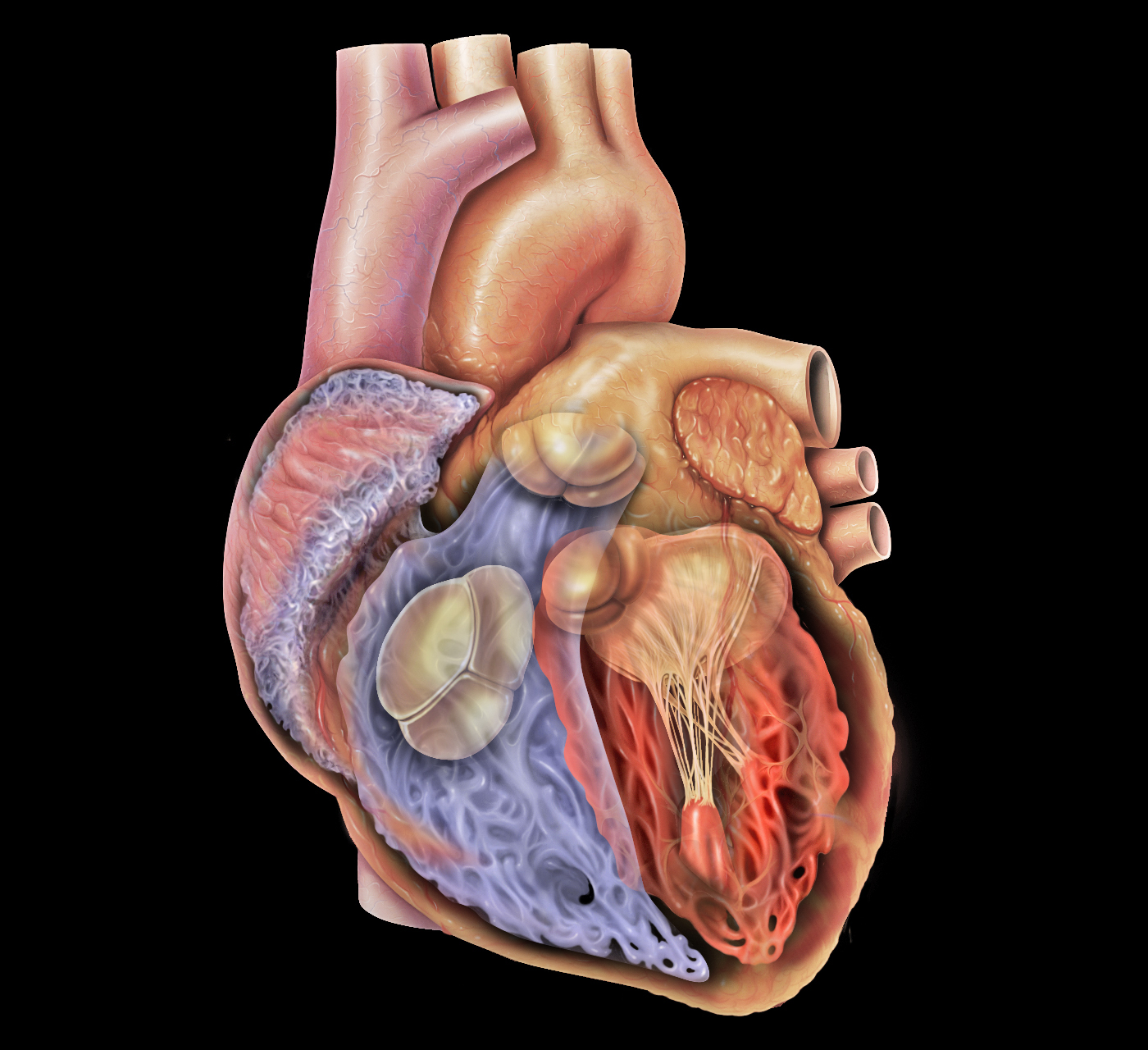
Anterior medical illustration: anatomical right is the viewer’s left. Attempt chamber, muscle, septum and valve identification before opening the key.
Check the identification key
In this anterior cutaway, anatomical right is the viewer’s left. Upper viewer-left: right atrium; lower viewer-left: right ventricle. Upper viewer-right: left atrium; lower viewer-right: left ventricle. Muscle surrounds the cavities; the muscular partition between the two lower cavities is the interventricular septum. Broad cusps at atrium–ventricle junctions are valve tissue; supporting cords below them are not the entire valve. This is a medical illustration, not a specimen photograph.
Use the image’s preparation and viewing direction
The image is a medical cutaway illustration by Patrick J. Lynch and C. Carl Jaffe, not a photograph of an intact heart. The front wall has been opened to reveal internal structures. Anatomical right is on the viewer’s left in this anterior view; the real structures have less regular shapes than the four-box model.
Locate the two upper atria and the two lower ventricles. The right atrium and right ventricle are on the viewer’s left; the left atrium and thick-walled left ventricle are on the viewer’s right. Identify the muscle as tissue around the cavities, not the hollow spaces containing blood.
Find the partition and the flaps
The interventricular septum runs between the ventricular cavities. Valve cusps lie at the atrium–ventricle junctions, with supporting cords projecting below them into the ventricles. Cords are not themselves valve flaps, and you should not label every thin internal strand as a valve.
Use the full-size image and then open the identification key after attempting the questions. This view is suitable for internal chamber, wall, septum and valve identification. It does not establish all coronary artery positions on the external surface; the separate coronary-image lesson supplies that comparison.
Sources for this lesson
Pause and explain
What separates the two ventricular cavities in this view?
Worked example
Identify the lower cavity on the viewer’s right and the thick tissue surrounding it.
Show the worked solution
- Translate viewer’s right to anatomical left.
- Separate the hollow cavity from its surrounding contractile tissue.
Answer The left ventricle is the cavity; the surrounding thick tissue is its muscular wall.
Try a topic test
Choose Easy, Normal or Difficult. Save your answers and open worked review after finishing.
Test this topic →From guided practice to a new situation
Write your answer and reasoning first. Use a hint only if you are stuck. The model solution is for self-checking; your written work is not automatically marked.
In the image, identify the upper receiving chamber and lower pumping chamber on the viewer’s left.
Give me a hint
This anterior view puts anatomical right on the viewer’s left.
Compare my reasoning
- Upper is right atrium.
- Lower is right ventricle.
Right atrium and right ventricle.
Look for these in your work
- Use correct anatomical side
- Distinguish receiving and pumping chambers
Identify the muscular wall and septum without using colour as your only evidence.
Give me a hint
Look around cavities and between the two lower cavities.
Compare my reasoning
- Outer surrounding tissue is muscular wall.
- The muscular partition between ventricles is the septum.
Muscular wall surrounds the cavities; the interventricular septum separates the two ventricular cavities.
Look for these in your work
- Use anatomical relationships
- Distinguish cavity from tissue
A learner labels a thin supporting cord below a valve cusp as the whole one-way valve. How should the identification improve?
Give me a hint
Compare the broad cusp with the cord attached below it.
Compare my reasoning
- The valve includes the cusp or flap at the junction.
- A supporting cord attaches to it but is not the whole valve.
Identify the cusp at the atrium–ventricle junction as valve tissue and distinguish its supporting cord.
Look for these in your work
- Locate the junction flap
- Avoid substituting a cord for a valve
Common mistakes
- Calling this image a photomicrograph.
- Labelling supporting cords as all the valve tissue.
What can you explain now?
What separates the two ventricular cavities in this view?
Compare with the explanation
Interventricular septum
The muscular partition lies between them.
After trying it yourself, choose your next review. This is your self-assessment.
Your review choice appears on Today. Sign in to sync it across devices.
Make it stick
Close the key and identify four chambers, wall, septum and valve tissue in the full-size image.
If you needed the solution, close it and solve the problem again from a blank page. A correct answer is stronger when you can explain the reason for each step.