Identify visible structures in a numbered flower photograph
Use the English key beside the unchanged published flower dissection.
Section 16.3 · Sexual reproduction in plants
Exam scope and assessment notes
Core and Extended · Biology 0970, 2026–2028. Use the stated specimen features and evidence. Diagrams are schematic. Written practice is self-checked; human subject-teacher review, supervised practical competence and cumulative assessment remain pending. Cambridge states that IGCSE 0970 has the same subject content as IGCSE 0610, with 9–1 grading. This StudyForA pathway maps the 2026–2028 syllabus only. This 9–1 graded qualification is available only in a limited number of administrative zones. Confirm that your centre can enter you for this code and exam series with Cambridge International. Cambridge has published a separate syllabus for 2029. StudyForA has not reconciled that version yet; this page supports exams in 2026, 2027 and 2028 only.
Before you begin
- Recall gametes, nuclear fusion, cellular respiration and the distinction between reproduction and growth.
Work on paper. Try each question before revealing help, and explain why your method works.
Quick readiness check
What is the source-key identity of 3?
What you will learn
- Translate the numbered key into anatomical names
- Connect visible parts without inventing hidden features
Identify visible structures in a numbered flower photograph
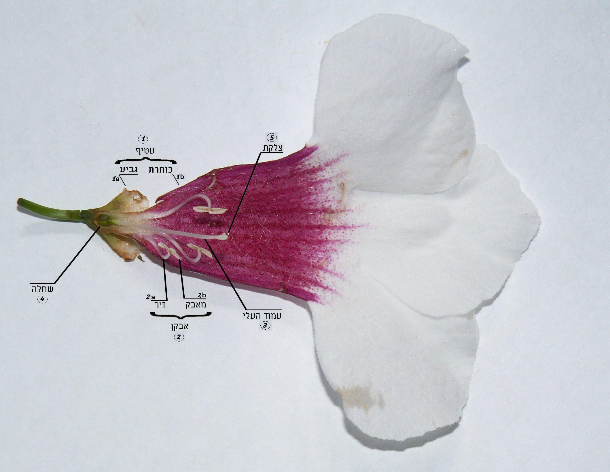
Unchanged numbered dissection: 1a sepals, 1b petals, 2 stamen, 2a filament, 2b anther, 3 style, 4 ovary, 5 stigma. Internal ovules are not visible. Original teaching data.
Numbered flower dissection

Enlarge image for inspection
Inspect the unchanged original at its native pixel size. Scroll inside the image to follow the structures.
Check the English identification key
- sepals: Outer brown/tan calyx tissue at the left base, aimed at by 1a. The calyx is the sepal group.
- petals: Broad white/magenta corolla tissue, aimed at by 1b. The corolla is the petal group.
- stamens: Group 2 brackets filament 2a and anther 2b. A filament plus its anther forms a stamen.
- filaments: Thin curved pale stalks supporting the elongated anthers; 2a points to a curled filament.
- anthers: Elongated pale pollen-bearing structures at the ends of filaments; 2b points to one.
- carpels: The pistil/carpel components are the connected stigma (5), style (3) and ovary (4). This is a grouping of visible components; no separate carpel label or carpel count is supplied.
- style: Central slender pale structure between the swollen green ovary and the enlarged stigma; 3 points to it.
- stigma: Enlarged pale tip of the central style, near the centre; 5 points to it.
- ovary: Swollen green structure at the left base; 4 points to it.
The ovary cavity and ovules are hidden in this view. The connected stigma, style and ovary are visible, but the number of constituent carpels is not established. The cut and spread corolla does not give a reliable original petal count. The photograph does not record a pollination event; its species is not specified.
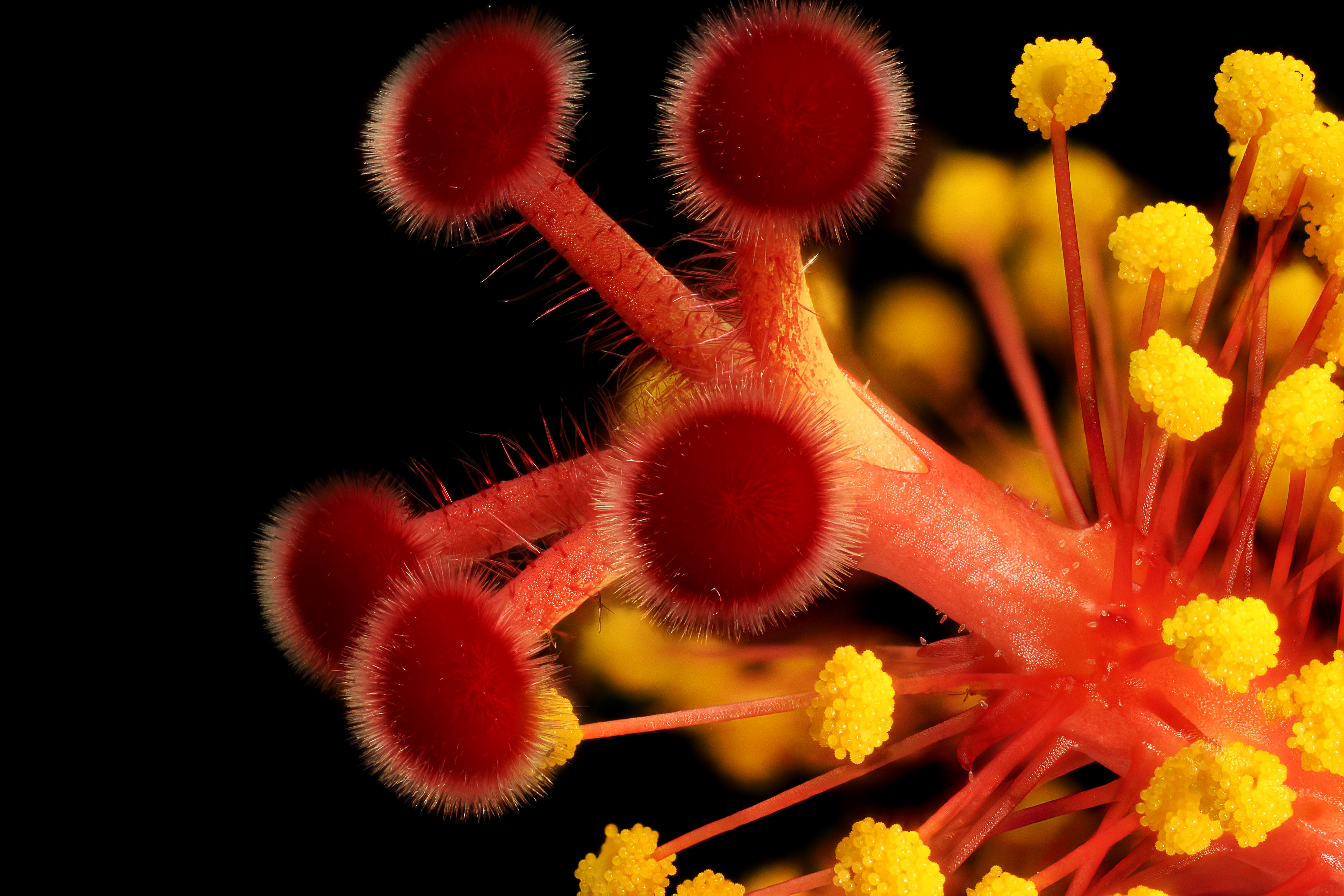
Stigma branches and stamens · hibiscus close-up

Enlarge image for inspection
Inspect the unchanged original at its native pixel size. Scroll inside the image to follow the structures.
Check the English identification key
- stigma: Large red hairy rounded heads on the left and centre.
- style: Exposed red branches immediately supporting those stigma heads.
- anthers: Yellow pollen-coated heads on the right and lower edge.
- filaments: Thin red stalks carrying those yellow anthers.
- stamens: Each yellow anther and its supporting thin red filament form one stamen.
- exclude staminal column: The thick main red central tube supports the many stamens and surrounds the style; it is the staminal column, not the exposed style branch.
Petals, sepals, ovary and ovules are outside the frame. The thick central staminal column is not the exposed style branch. Use the exposed branches immediately supporting the red stigma heads for style identification. The photograph does not record an insect visit or a pollination event.
| Feature | Meaning |
|---|---|
| Outer groups | 1a sepals; 1b petals |
| Stamen | 2 includes filament 2a and anther 2b |
| Carpel components | Stigma 5, style 3 and ovary 4 |
| Limit | No visible ovules, carpel count or recorded pollination |
Translate the numbered key into anatomical names
The unchanged published photograph shows a dissected flower, rather than an intact flower seen from the front. Its original labels use Hebrew and numbers. The surrounding English key identifies 1a as the calyx or sepal group, 1b as the corolla or petal group, and 2 as the stamen grouping. Within 2, the slender curved supporting stalk at 2a is a filament and the elongated pale structure at 2b is an anther.
The central reproductive structure connects the enlarged stigma at 5 to the swollen green ovary at 4 through the slender style at 3. Stigma, style and ovary are carpel components. The photograph calls their combined structure a pistil, but it does not establish how many constituent carpels are present or whether carpels have fused. Naming the visible components is stronger evidence than claiming an unsupported carpel count.
Connect visible parts without inventing hidden features
Use connections, leader endpoints and the source key together. The tan basal tissue at 1a is a sepal group in this specimen; sepals need not always be green. The spread white and magenta corolla at 1b is petal tissue, but cutting and spreading it prevents a confident original petal count. The image does not document a species or an insect visit, so those are not facts to infer from its attractive colours.
Although dissection reveals external parts clearly, this view does not expose the ovary cavity or its ovules. Enlargement helps inspect recorded detail; it does not make the ovary transparent. Connect supported identities with functions: anthers produce pollen, filaments support them, the stigma receives pollen and the ovary contains ovules. Use the separate cut-section photograph for internal ovule identification. The accompanying hibiscus close-up supplies another real-image check: red stigma heads and exposed style branches differ from the yellow anthers on thin filaments. Its thick central staminal column must not be labelled style, and its ovary and outer flower parts are outside the frame.
Pause and explain
Which internal feature cannot be located in this photograph?
Worked example
Use labels 2a and 2b to identify the two parts and their complete unit.
Show the worked solution
- 2a points to the supporting filament.
- 2b points to the anther; together they form a stamen.
Answer 2a filament; 2b anther; complete unit stamen.
Try a topic test
Choose Easy, Normal or Difficult. Save your answers and open worked review after finishing.
Test this topic →From guided practice to a new situation
Write your answer and reasoning first. Use a hint only if you are stuck. The model solution is for self-checking; your written work is not automatically marked.
Name 1a, 1b, 2a and 2b using the English photograph key.
Give me a hint
Separate outer groups from the stamen components.
Compare my reasoning
- 1a is the sepal group and 1b the petal group.
- 2a is filament and 2b anther.
1a sepals; 1b petals; 2a filament; 2b anther.
Look for these in your work
- Match each number to the supported feature
- Keep group and component names distinct
Name 3, 4 and 5, and describe how their functions connect.
Give me a hint
Follow the central structure from receiving tip to basal organ.
Compare my reasoning
- 5 receives pollen; 3 provides the connecting tube route.
- 4 encloses ovules, although those ovules are hidden here.
3 style; 4 ovary; 5 stigma. The stigma receives pollen, the style provides the pollen-tube route toward the ovary, and the ovary contains ovules.
Look for these in your work
- Assign all three labels
- Connect identities with functions
Does this dissection prove a precise carpel count, visible ovules or a successful insect pollination event?
Give me a hint
Separate the photographed anatomy from unavailable evidence.
Compare my reasoning
- The key identifies carpel components but not their constituent count.
- Internal ovules and transfer events are not recorded.
No. Identify the visible stigma, style and ovary; use an internal view for ovules and separate evidence for carpel count or pollination.
Look for these in your work
- Retain the actual visible evidence
- State all three unsupported inferences
Common mistakes
- Calling the entire central structure one egg cell.
- Inventing ovules or a carpel count from the external view.
What can you explain now?
Which internal feature cannot be located in this photograph?
Compare with the explanation
Ovules
The ovary cavity is not exposed.
After trying it yourself, choose your next review. This is your self-assessment.
Your review choice appears on Today. Sign in to sync it across devices.
Make it stick
Identify supported photographic parts and explain why the internal ovules require another view.
If you needed the solution, close it and solve the problem again from a blank page. A correct answer is stronger when you can explain the reason for each step.