Identify glands in the published ovarian anatomy image
Trace adrenal, pancreatic and ovarian leaders in the unchanged Blausen image.
Section B13.2 · Hormones
Exam scope and assessment notes
Mapped to the named 0973 section using teaching from the corresponding single-science course. A 0973-specific review of every Core and Supplement objective is still in progress. Written practice is self-checked; it does not demonstrate supervised laboratory competence or award an official Cambridge grade. This qualification reports two grades on the 9–1 scale; confirm the entry route for your zone and exam series.
Before you begin
- Recall electrical impulses, blood transport, gland secretion and the distinction between a stimulus and a body response.
Work on paper. Try each question before revealing help, and explain why your method works.
Quick readiness check
What does Gonads (Ovaries) identify here?
What you will learn
- Read the anterior and posterior source views
- Supply hormone names from the teaching
Identify glands in the published ovarian anatomy image
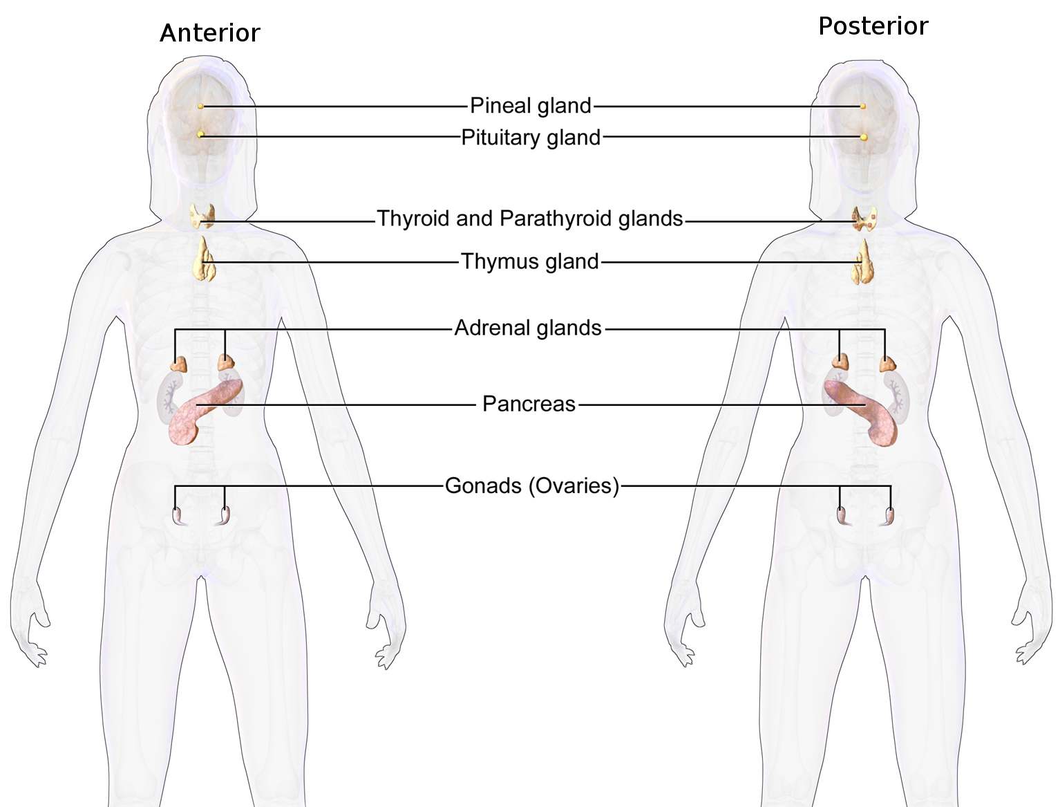
Unchanged published ovarian anatomical illustration: anterior left, posterior right. Adrenal caps lie above kidneys; pancreas spans upper abdomen; ovarian leaders branch within pelvis. Testes are absent.
Enlarge image for inspection
Pan across the source at its native image size to follow the leaders. Try identification before opening the key.
Check the identification key
Adrenal glands: paired small brown caps above kidneys → adrenaline. Pancreas: pink elongated upper-abdominal gland → insulin. Gonads (Ovaries): paired pelvic ovaries → oestrogen. Testes are absent; use the companion testicular source.
Read the anterior and posterior source views
The published Endocrine System (Female) illustration by BruceBlaus and Blausen Medical Communications shows anterior and posterior views side by side. Transparent body outlines reveal internal glands. These are anatomical illustrations, not photographs or section planes, and their colours and proportions are schematic. Begin by locating the abdomen and pelvis in each view.
The Adrenal glands label branches towards both small brown caps above the larger pale kidneys in each view. Pancreas has a leader ending inside the pink elongated upper-abdominal gland. Gonads (Ovaries) branches to the two small ovarian structures within the pelvis. Read the actual line endpoints rather than identifying a gland from the nearest printed word.
Supply hormone names from the teaching
Add the required hormone pairings yourself: adrenal glands secrete adrenaline, pancreas secretes insulin, and ovaries secrete oestrogen. The source prints gland names but not these hormone names. Testes and testosterone are the fourth required Core pairing, covered by the companion testicular image rather than by inventing a testis in this ovarian source.
Extra head, neck and chest glands are visible but exceed this four-gland identification task. The image does not distinguish hormone-producing cell populations or measure secretion. Enlarge the unchanged source to follow its leaders and attempt identification before opening the separate key. The credited historical licence applies to this exact unchanged source version.
Sources for this lesson
Pause and explain
Which interpretation follows the actual adrenal leaders?
Worked example
Identify the three required glands visible in the source and give their hormones.
Show the worked solution
- Adrenal leaders reach caps; pancreas reaches the elongated abdominal gland.
- Gonads (Ovaries) reaches paired pelvic ovaries.
Answer Adrenal glands–adrenaline; pancreas–insulin; Gonads (Ovaries)–oestrogen. Testes are absent from this source.
Try a topic test
Choose Easy, Normal or Difficult. Save your answers and open worked review after finishing.
Test this topic →From guided practice to a new situation
Write your answer and reasoning first. Use a hint only if you are stuck. The model solution is for self-checking; your written work is not automatically marked.
Identify all three required visible source glands and state their hormones.
Give me a hint
Read the three abdominal and pelvic leaders.
Compare my reasoning
- Adrenal glands/adrenaline and pancreas/insulin.
- Gonads (Ovaries) identifies ovaries/oestrogen.
Adrenal glands–adrenaline; pancreas–insulin; ovaries–oestrogen.
Look for these in your work
- Trace all three targets
- Supply the unprinted hormone names
Explain how to distinguish the Adrenal glands and Pancreas endpoints in this image.
Give me a hint
Compare caps above kidneys with the elongated organ.
Compare my reasoning
- Adrenal branches end at small brown caps above the kidneys.
- Pancreas ends inside the pink elongated upper-abdominal gland.
Adrenal glands are the small caps above kidneys; Pancreas is the separate pink elongated upper-abdominal organ. Their hormones are adrenaline and insulin respectively.
Look for these in your work
- Describe both endpoints
- Match their hormones
A learner identifies the fourth required gland as testes within this source. Correct the identification and say where to find that fourth pairing.
Give me a hint
This source shows ovaries, not testes.
Compare my reasoning
- The pelvic Gonads (Ovaries) targets secrete oestrogen.
- The companion testicular source shows testes, which secrete testosterone.
This source shows ovaries–oestrogen. Testes–testosterone is identified in the separate companion testicular image.
Look for these in your work
- Correct the actual source target
- Locate the missing required pair
Common mistakes
- Naming the kidneys at the adrenal leader endpoints.
- Treating the printed gland names as printed hormone names.
What can you explain now?
Which interpretation follows the actual adrenal leaders?
Compare with the explanation
Small brown caps above the kidneys; adrenaline
The caps are adrenal glands.
After trying it yourself, choose your next review. This is your self-assessment.
Your review choice appears on Today. Sign in to sync it across devices.
Make it stick
Trace the three required source targets and identify the missing companion pairing.
If you needed the solution, close it and solve the problem again from a blank page. A correct answer is stronger when you can explain the reason for each step.