Recognise lymphocytes and phagocytes in photomicrographs
Compare source-confirmed columns F and G in genuine stained blood images.
Section 9.4 · Blood
Extended only (Supplement); Biology 0610, 2026–2028. Use the stated specimen features and evidence. Diagrams are schematic. Written practice is self-checked
Before you begin
- Recall the two white-cell diagram examples.
Work on paper. Try each question before revealing help, and explain why your method works.
Quick readiness check
What evidence confirms the column identities?
What you will learn
- Find the two required columns in the whole figure
- Use verified identity with visible morphology
Photomicrographs: focus on F and G
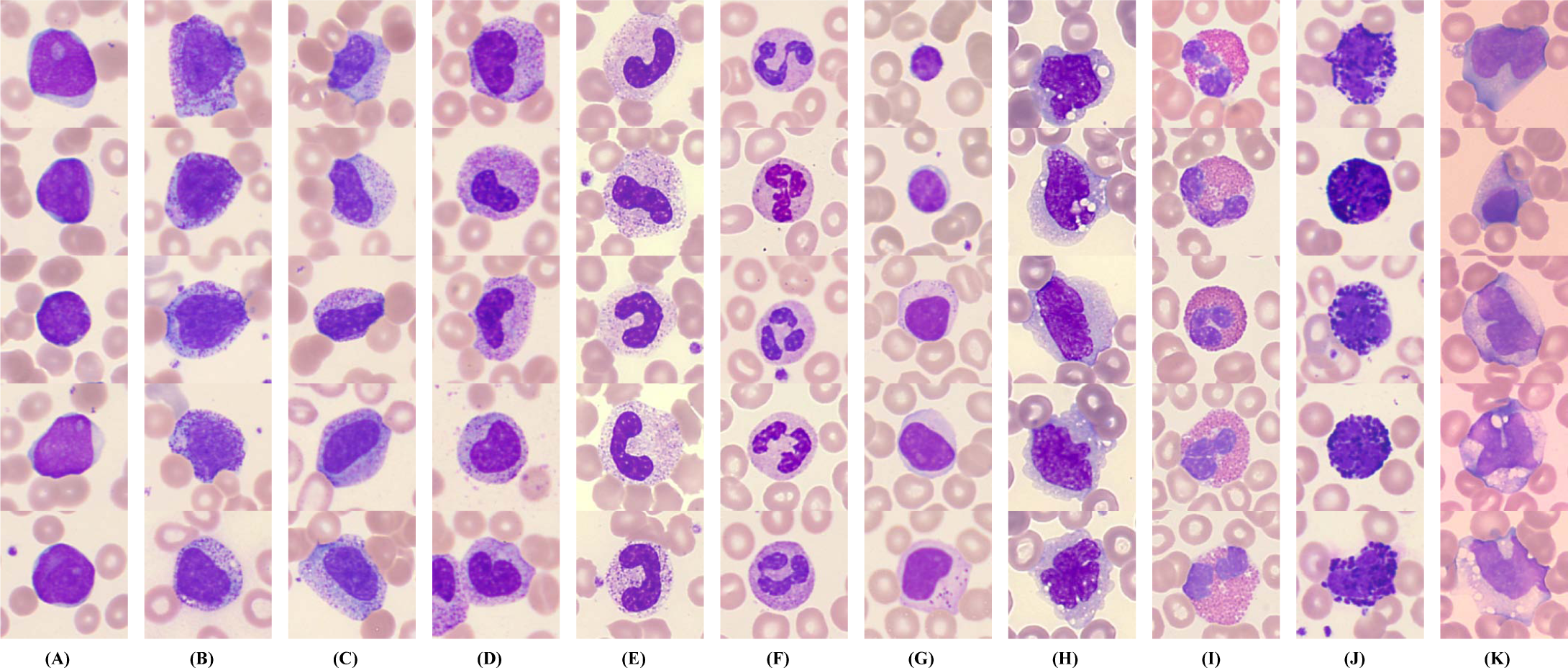
F contains source-confirmed segmented neutrophils, a phagocyte type; G contains lymphocytes. Other columns are outside this comparison, and selected tiles are not a blood count.
Enlarge image for inspection
Scroll across the enlarged image to inspect labels and cell features. The whole source image is retained. Use the identification key after trying the questions.
Check the identification key
Column F: segmented neutrophils, a phagocyte type, with lobed nuclei and visible cytoplasm. Column G: lymphocytes, with a large round nucleus occupying much of each cell and a relatively narrow rim of cytoplasm. These identities are confirmed by the publisher’s Figure 2 legend, not inferred from purple colour alone. Other columns illustrate additional cell categories beyond this IGCSE comparison. Images are examples, not a blood-count sample or a diagnostic task; the whole figure is unchanged and no common calibrated scale is supplied for comparing absolute cell sizes.
Find the two required columns in the whole figure
The supplied unchanged Figure 2 from Wang and colleagues contains genuine stained peripheral-blood photomicrographs in columns A to K. This IGCSE task uses F and G. The publisher identifies F as segmented neutrophils and G as lymphocytes. Neutrophils are phagocytes, so F provides the required phagocyte examples.
Column F shows lobed or segmented nuclei with visible cytoplasm. Column G shows large round nuclei occupying much of each cell, with comparatively narrow cytoplasm rims. Compare several clear examples within each column; variability and preparation mean that no single colour patch is a complete definition.
Use verified identity with visible morphology
The source legend confirms identities, while morphology explains why these examples fit the teaching comparison. Purple is a stain colour shared by many nuclei and fragments, so it cannot distinguish lymphocyte from phagocyte by itself. Other columns include cell categories outside this IGCSE exercise; they are retained because the whole source figure is unchanged.
Open the full-size image to read the column letters before answering, then reveal the key. There is no common calibrated scale for calculating or comparing absolute sizes across these image tiles. These selected examples are not a representative differential blood count and should not be used to diagnose a patient or the learner.
Pause and explain
Which columns supply the required comparison?
Worked example
Identify F and G at the IGCSE phagocyte/lymphocyte level and give one visible feature for each.
Show the worked solution
- F is source-confirmed segmented neutrophils, a phagocyte type.
- G is source-confirmed lymphocytes with large round nuclei.
Answer F phagocyte examples with lobed nuclei; G lymphocytes with large round nuclei and narrower cytoplasm rims.
Try a topic test
Choose Easy, Normal or Difficult. Save your answers and open worked review after finishing.
Test this topic →From guided practice to a new situation
Write your answer and reasoning first. Use a hint only if you are stuck. The model solution is for self-checking; your written work is not automatically marked.
Find F in the whole figure and name the IGCSE category and source-confirmed type.
Give me a hint
Read the column letter before comparing nuclei.
Compare my reasoning
- F is labelled segmented neutrophils by the publisher.
- Neutrophils are phagocytes.
F: phagocyte examples, specifically segmented neutrophils.
Look for these in your work
- Locate F
- Name category and example
Find G and describe two features supporting lymphocyte recognition.
Give me a hint
Compare nucleus occupancy and cytoplasm.
Compare my reasoning
- G has a large round nucleus.
- A relatively narrow cytoplasm rim remains.
G lymphocytes: large round nucleus and narrow cytoplasm rim.
Look for these in your work
- Locate G
- Give both features
Why would counting all F and G tiles not establish a patient’s phagocyte-to-lymphocyte ratio?
Give me a hint
The figure selects examples of categories.
Compare my reasoning
- Tiles are curated illustrations of cell classes.
- They are not a representative measured sample.
The curated figure is for recognition, not a representative blood count, so its tile totals cannot establish a patient’s ratio.
Look for these in your work
- Distinguish selected examples from sampling
- Reject the clinical inference
Common mistakes
- Choosing from purple colour alone.
- Treating curated image columns as a measured blood count.
What can you explain now?
Which columns supply the required comparison?
Compare with the explanation
F phagocytes; G lymphocytes
F contains segmented neutrophils.
After trying it yourself, choose your next review. This is your self-assessment.
Your review choice appears on Today. Sign in to sync it across devices.
Make it stick
Identify F and G in the full-size figure and explain the evidence limits.
If you needed the solution, close it and solve the problem again from a blank page. A correct answer is stronger when you can explain the reason for each step.