Identify incisors, canines, premolars and molars
Use position and crown shape to identify adult tooth types.
Cross-referenced to the 2028–2029 0653 section using original teaching from the related single-science courses. A 0653-specific objective-by-objective and Core/Supplement review is in progress. Written practice is self-checked and does not demonstrate supervised laboratory competence or award an official Cambridge grade.
Before you begin
- Recall large and small nutrient molecules, cell membranes and enzyme specificity.
- Use the stated diagram keys and evidence; models are not to scale.
Work on paper. Try each question before revealing help, and explain why your method works.
Quick readiness check
Which teeth lie at the front with cutting edges?
What you will learn
- Use position and crown shape to identify adult tooth types.
- Explain which features an external photograph cannot reveal.
Identify a permanent dentition
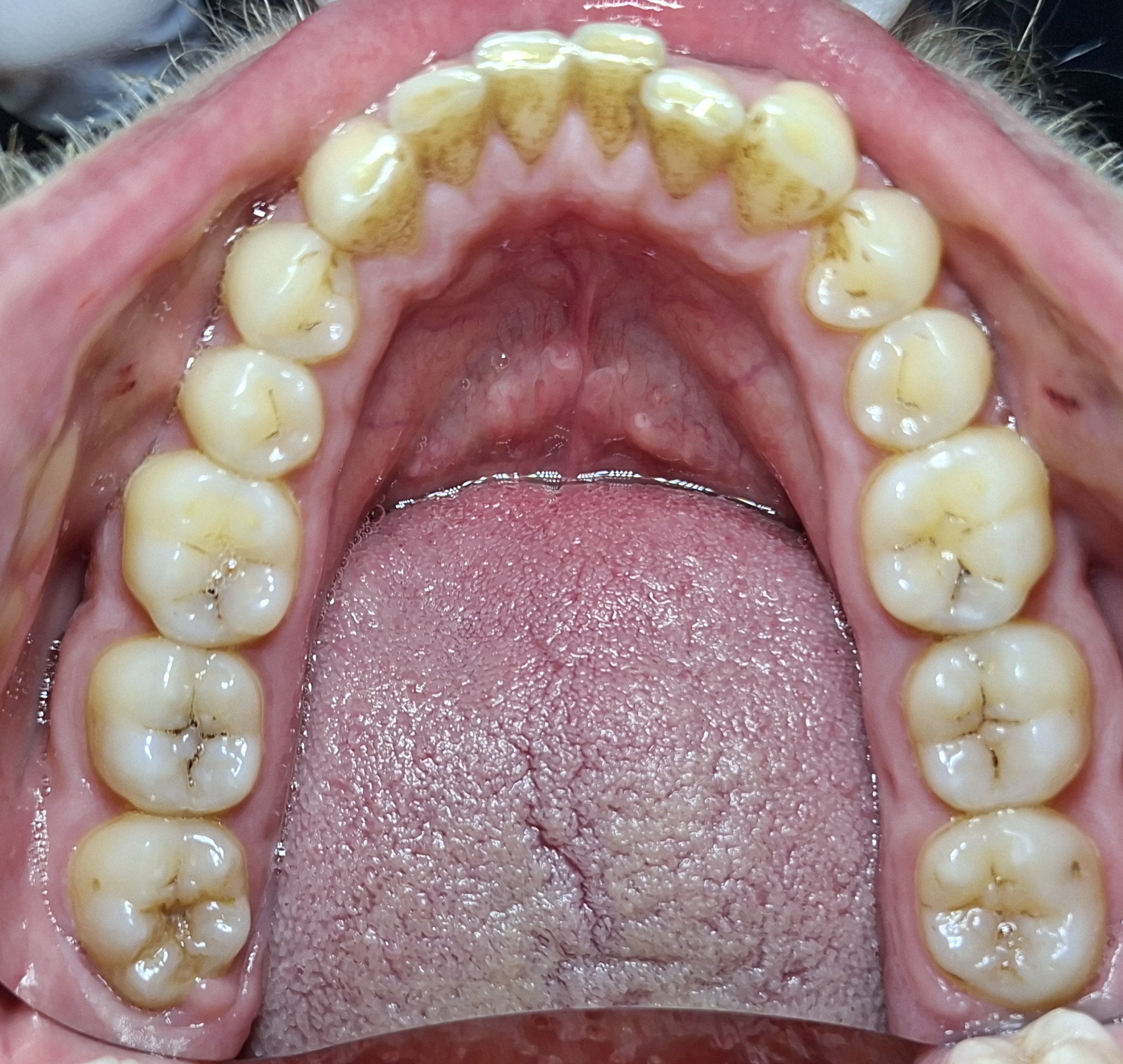
The genuine lower-teeth photograph shows incisors at the front, canines beside them, premolars behind and molars farther back. Surface staining is visible. Use position and crown shape, rather than diagnosing oral health from the photograph.
Check the identification key
Begin at the front centre near the top of the image: the four edged front teeth are incisors. Moving toward the back on each side, the next pointed tooth is a canine, followed by two premolars and then broader molars. The photograph is a permanent dentition; this pattern does not establish a universal count for every age or person. Internal enamel, dentine and pulp are not resolved by the outside view.
Four types in a permanent dentition
Incisors are front teeth with cutting edges. Canines lie beside them and have pointed crowns. Premolars are behind canines with broader cusped surfaces; molars lie farther back and have broad grinding surfaces.
Use position and shape together. Premolars and molars can both have cusps, so a cusp alone does not uniquely identify a tooth. Tooth number and visibility vary between people and developmental stages.
Read photographs carefully
The supplied public-domain photograph shows a lower permanent dentition from above. Starting at the front centre, identify incisors, then canines, premolars and molars moving toward the back on each side. The tongue is not another tooth.
This photograph shows surface features rather than the internal enamel/dentine/pulp layers. Staining is visible, but the picture is used for identification, not to diagnose an individual’s oral health.
Sources for this lesson
Pause and explain
Which feature best supports canine identification?
Worked example
A tooth is immediately behind a canine and has a broad cusped crown. Which type is best supported?
Show the worked solution
- Use the stated position.
- Combine it with the crown shape.
- Identify a premolar.
Answer A premolar; position behind the canine supports the identification.
Try a topic test
Choose Easy, Normal or Difficult. Save your answers and open worked review after finishing.
Test this topic →From guided practice to a new situation
Write your answer and reasoning first. Use a hint only if you are stuck. The model solution is for self-checking; your written work is not automatically marked.
Name the four required tooth types in order from front toward back.
Give me a hint
Start with cutting teeth.
Compare my reasoning
- Incisors then canines.
- Premolars then molars.
Incisors → canines → premolars → molars.
Look for these in your work
- All four.
- Correct general adult order.
In the supplied photograph, describe where to find premolars relative to canines and molars.
Give me a hint
Move from front to back.
Compare my reasoning
- Behind canines.
- In front of molars.
Premolars lie between canines and molars on each side.
Look for these in your work
- Both neighbours.
- Use position rather than colour.
An isolated tooth has a cusped crown but its position is unknown. Can “cusped” alone prove it is a premolar? Explain.
Give me a hint
Molars also have cusps.
Compare my reasoning
- Crown features overlap.
- Position or more structural evidence is needed.
No; molars also have cusps, so more identifying evidence is needed.
Look for these in your work
- Recognise shared feature.
- Limit certainty.
Common mistakes
- A cusp alone does not uniquely identify a tooth.
- An adult pattern is not a universal count for children.
What can you explain now?
Which feature best supports canine identification?
Compare with the explanation
A pointed crown beside the incisors.
Use shape and position together.
After trying it yourself, choose your next review. This is your self-assessment.
Your review choice appears on Today. Sign in to sync it across devices.
Make it stick
Identify the four tooth types using shape and position together.
If you needed the solution, close it and solve the problem again from a blank page. A correct answer is stronger when you can explain the reason for each step.