Use microscope images without inventing invisible structures
Compare real onion, cheek and bacterial images with labelled models.
Cross-referenced to the 2028–2029 0653 section using original teaching from the related single-science courses. A 0653-specific objective-by-objective and Core/Supplement review is in progress. Written practice is self-checked and does not demonstrate supervised laboratory competence or award an official Cambridge grade.
Before you begin
- Recall the distinction between plant, animal and bacterial cells. Read the supplied evidence before drawing a conclusion.
Work on paper. Try each question before revealing help, and explain why your method works.
Quick readiness check
Which supplied image shows bacterial external surfaces?
What you will learn
- Identify visible cell boundaries and nuclei where supported.
- Separate observed features from model-based expectations.
Real images have visibility limits
Onion epidermis · light microscope
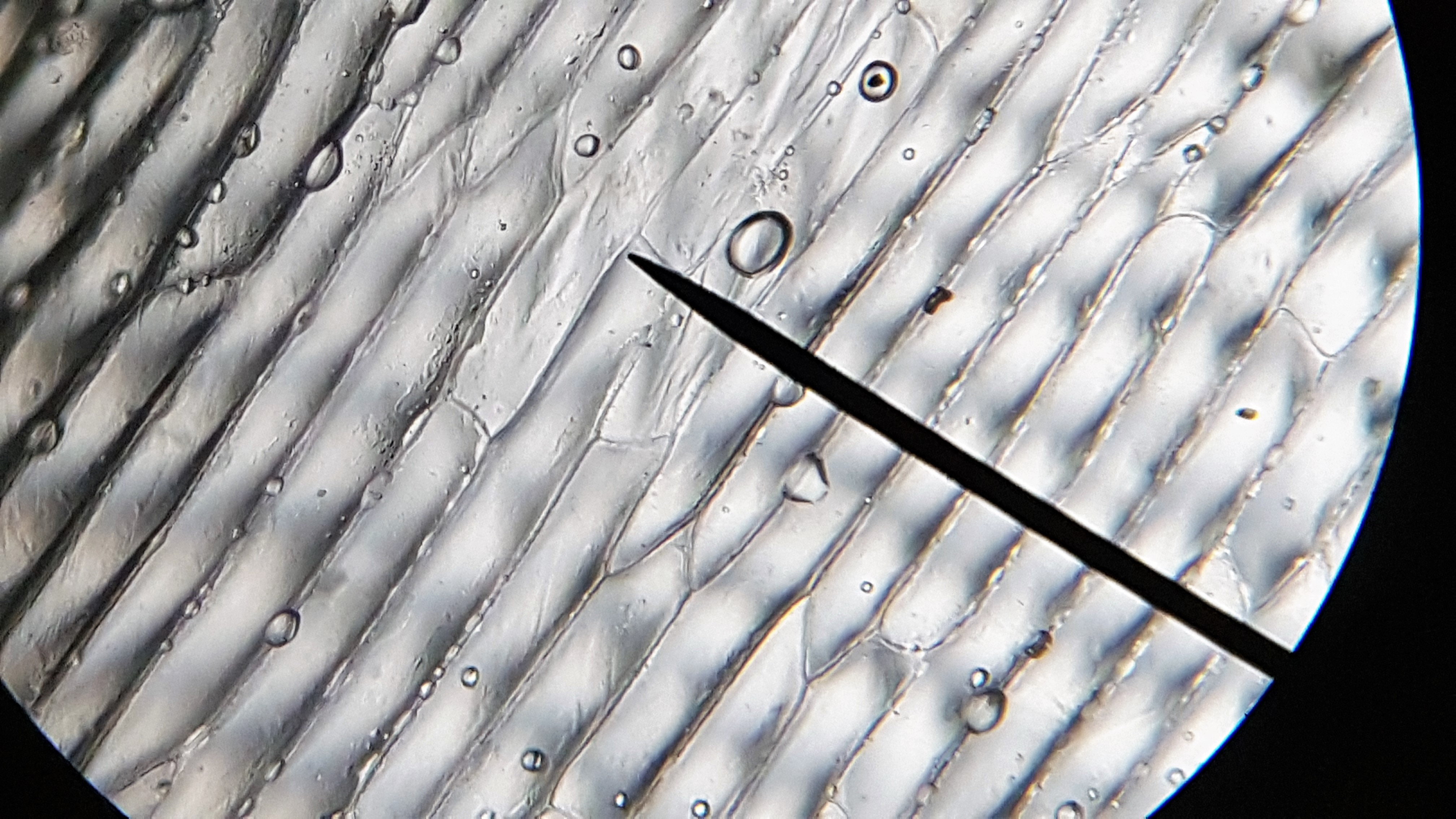
Photo: SaraKasa · CC BY 4.0 · Original image unchanged; displayed responsively.
Stained cheek cells · light microscope
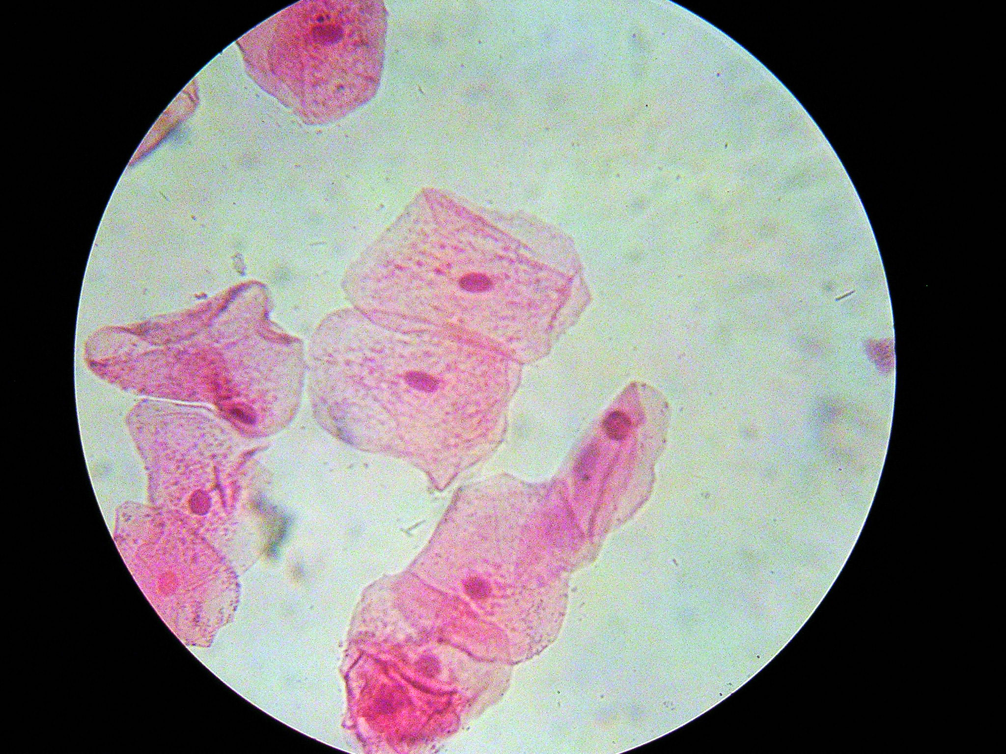
Photo: Mulletsrokk · CC BY-SA 3.0 · Original image unchanged; displayed responsively.
E. coli · scanning electron microscope
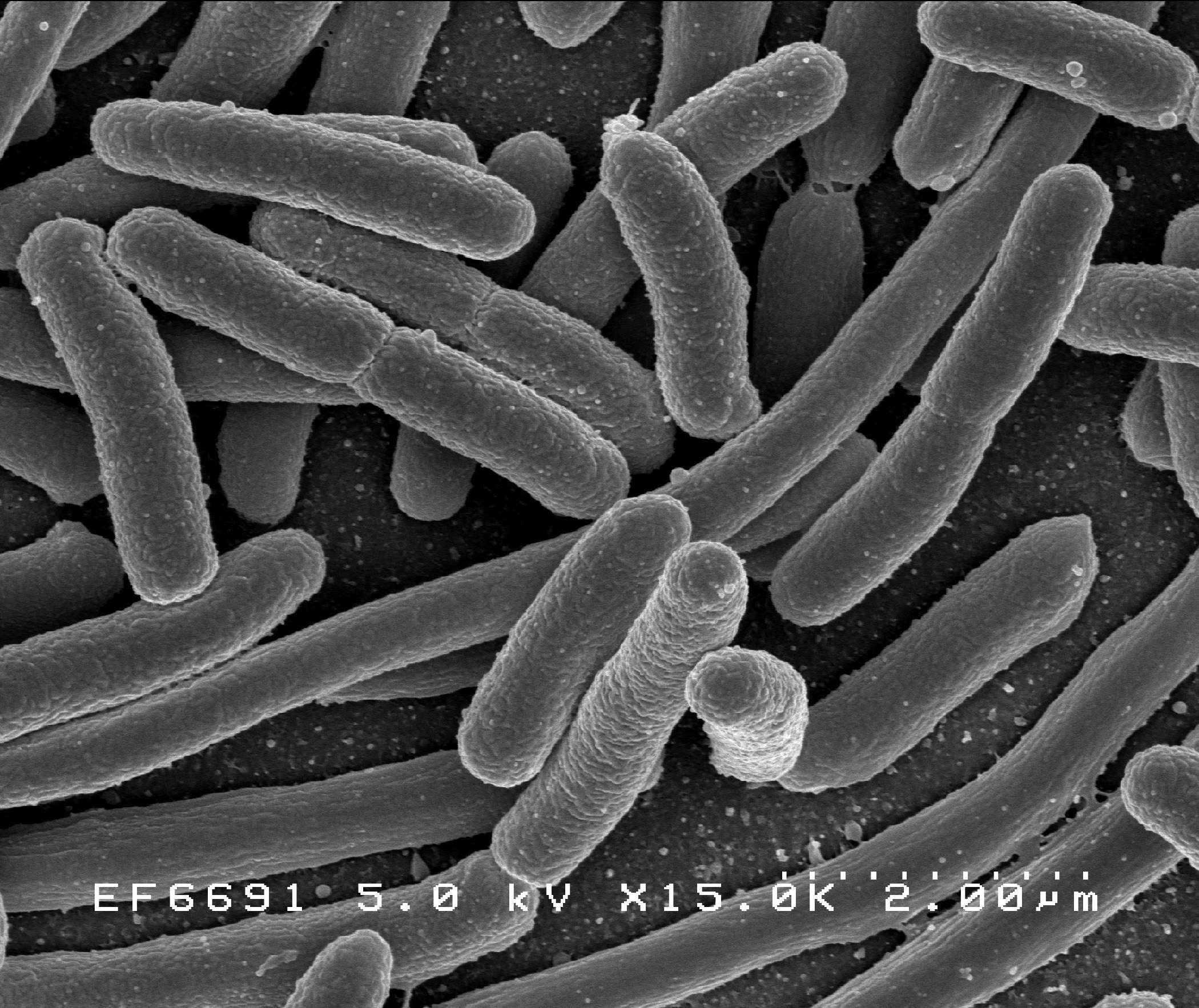
Photo: NIAID / Rocky Mountain Laboratories, NIH · CC BY 2.0 · Original image unchanged; displayed responsively.
Compare observed image features with the labelled cell models. These photographs support interpretation practice, not certification of supervised microscope competence.
Observe before naming
The onion epidermis image shows elongated adjoining cells with conspicuous wall boundaries. Small circular bright objects may be preparation artefacts or bubbles: they should not be labelled automatically as nuclei or chloroplasts. Nuclei are not resolved clearly in this supplied onion image. Bulb epidermis is non-photosynthetic and does not require visible chloroplasts.
The stained cheek-cell image shows irregular cell outlines, paler cell contents and darker rounded nuclei. These are animal cells without cellulose walls. The outline locates the cell boundary, but the thickness of the membrane itself is below what this photograph resolves.
A surface view has a different limit
The scanning electron micrograph shows rod-shaped E. coli cells and their external surfaces. It does not reveal the internal circular DNA, plasmids, ribosomes or cytoplasm. Use the bacterial schematic for those internal labels rather than placing them on an opaque surface photograph.
Staining, focus, magnification and resolution affect visibility. Enlarging a blurred picture makes the blur larger; it does not recover missing detail. Compare several cells, avoid overlapping regions and distinguish visible evidence from a structure expected from biological knowledge. These supplied-image exercises do not certify supervised microscope competence.
Sources for this lesson
Pause and explain
What is a defensible claim about the supplied onion image?
Worked example
In the supplied cheek image, what supports a nucleus label, and why should ribosomes not be labelled?
Show the worked solution
- Darker rounded regions lie within the cell outlines.
- The specimen is a stained cheek-cell preparation.
- Individual ribosomes are not resolved in this image.
Answer Dark internal rounded regions support nuclei; ribosomes are not resolved.
Try a topic test
Choose Easy, Normal or Difficult. Save your answers and open worked review after finishing.
Test this topic →From guided practice to a new situation
Write your answer and reasoning first. Use a hint only if you are stuck. The model solution is for self-checking; your written work is not automatically marked.
Describe one visible plant feature in the onion image and one structure not clearly resolved.
Give me a hint
Use the supplied image, not a memorised coloured model.
Compare my reasoning
- Adjoining wall boundaries are conspicuous.
- Nuclei or individual mitochondria are not clearly resolved.
Visible walls; nuclei or mitochondria not clearly resolved.
Look for these in your work
- Name a visible boundary.
- State a genuine visibility limit.
Compare a darker internal region in a cheek cell with a rod outline in the bacterial surface image.
Give me a hint
The images provide different kinds of evidence.
Compare my reasoning
- The cheek region supports a nucleus identification.
- The rod outline identifies a bacterial external boundary, not its DNA.
Cheek: nucleus evidence; bacterium: surface outline, not an internal DNA label.
Look for these in your work
- Keep the two image types distinct.
- Avoid invented internal labels.
A student labels ribosomes on the onion image by adding dots that are not visible. Propose a better answer.
Give me a hint
Expected structures may be explained separately.
Compare my reasoning
- State ribosomes are expected from the cell model.
- State they are not resolved in this photograph.
- Use a suitable higher-resolution image or the labelled schematic for identification.
Separate expected ribosomes from visible evidence; do not add invented observed dots.
Look for these in your work
- Distinguish expectation and observation.
- Suggest suitable evidence.
Common mistakes
- Unseen does not necessarily mean absent.
- Do not mistake bubbles or overlap for organelles.
What can you explain now?
Does enlarging an unresolved image prove a previously invisible organelle is absent?
Compare with the explanation
No; resolution limits visibility.
Expected cell structures and visible evidence are different claims.
After trying it yourself, choose your next review. This is your self-assessment.
Your review choice appears on Today. Sign in to sync it across devices.
Make it stick
Compare one visible feature and one unresolved structure in each supplied image.
If you needed the solution, close it and solve the problem again from a blank page. A correct answer is stronger when you can explain the reason for each step.