Recognise gas-exchange tissues in images
Use visible tissue architecture to identify airway and alveolar tissues in slides and micrographs.
AS Level; Cambridge International Biology 9700, 2028–2030. Written practice is self-checked; microscope recognition, biological drawing and supervised practical competence require practical work, while human subject-teacher review remains pending.
Before you begin
- Know that gas exchange is diffusion across a surface and that ventilation renews air while blood flow renews the blood side.
Work on paper. Try each question before revealing help, and explain why your method works.
Quick readiness check
What visible feature supports identifying ciliated epithelium?
What you will learn
- Recognise cartilage, ciliated and squamous epithelium, goblet cells, smooth muscle and capillaries
- Support identifications using visible evidence rather than an assumed label
Identify major respiratory organs in a published image
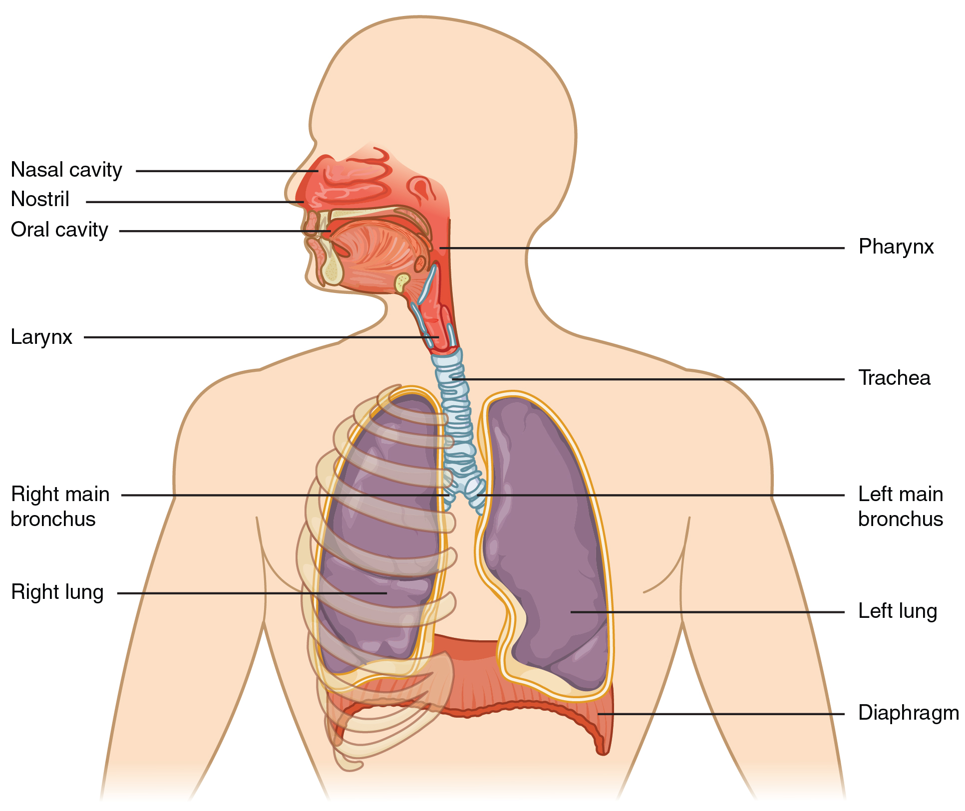
Published anatomical illustration: identify the larger airways, paired lungs and diaphragm. Use the enlargement for labels; this is not a clinical image.
Enlarge image for inspection
Scroll across the whole source image to inspect its labels. Try identification before opening the key.
Check the identification key
lungs: The two large purple organs on either side of the central airway; patient's right is image left. diaphragm: Red domed sheet beneath both lungs, indicated by the lower-right label. larynx: The labelled red and blue cartilaginous region above the ringed trachea. trachea: Vertical pale-blue ringed airway down the centre of the neck and chest. bronchi: Two branches at the bottom of the trachea entering the lungs, identified by Right main bronchus and Left main bronchus labels. ribs: Translucent curved beige bones drawn across the right lung on image left. Use the thorax figure for an explicit Ribs label. No bronchioles, alveoli, capillaries or intercostal labels. Ribs are drawn but not labelled. Anatomical left/right is the person's left/right, opposite the viewer.
Build the explanation
Cartilage appears as a firm supporting tissue within larger airway walls. Ciliated epithelial cells form a lining with hair-like cilia at the lumen surface; goblet cells appear among the lining cells and may contain visible secretory material. Smooth muscle forms bands or layers of elongated cells within airway walls. Alveolar squamous cells are very thin, and the air spaces are separated by delicate septa with capillaries close by. A micrograph may show only part of a cell or wall, so the image scale and context matter.
Apply it carefully
Identify a tissue with two clues where possible: location plus shape, or lining plus associated structure. A stain can make features easier to see but colour is not universal evidence. Distinguish directly visible features from the expected anatomy of a labelled diagram; a written exercise does not certify hands-on microscopy.
Pause and explain
What evidence supports identifying an alveolar capillary?
Worked example
A lining has a free surface with many short projections into an airway lumen. What feature is visible?
Show the worked solution
- The projections are on the epithelial surface.
- They are consistent with cilia.
Answer Cilia on ciliated epithelial cells.
Try a topic test
Choose Easy, Normal or Difficult. Save your answers and open worked review after finishing.
Test this topic →From guided practice to a new situation
Write your answer and reasoning first. Use a hint only if you are stuck. The model solution is for self-checking; your written work is not automatically marked.
Give two clues that support identifying a ciliated epithelial lining in an airway micrograph.
Give me a hint
Use the lumen surface and the cells forming the lining.
Compare my reasoning
- The cells form an epithelial sheet facing the lumen.
- Short cilia project from the free surface.
The lining cells form an epithelial layer facing the airway lumen, and cilia project from their free surface. Use the image scale and airway context to support the identification.
Look for these in your work
- Lining position stated
- Cilia identified
- Context used
A tiny blood-filled channel follows a thin wall around an air space. What two structures are likely visible?
Give me a hint
Identify the air space boundary and the vessel beside it.
Compare my reasoning
- The air space is an alveolus.
- The nearby channel is a capillary in the alveolar septum.
The air space is an alveolus and the small blood-filled channel beside its thin wall is a capillary in the septum.
Look for these in your work
- Alveolus identified
- Capillary identified
- Relationship stated
An image is stained dark and the cell boundaries are unclear. Explain how to report a tentative tissue identification responsibly.
Give me a hint
Separate confidence from evidence.
Compare my reasoning
- Describe the visible position and shape first.
- State which features are indistinct and avoid claiming an unsupported label.
Describe the visible evidence and image context, such as a lining facing a lumen or a thin septum beside a small vessel. State that stain or resolution obscures boundaries and make a cautious identification rather than treating an expected label as observed fact.
Look for these in your work
- Evidence described
- Image limitation acknowledged
- Inference calibrated
Common mistakes
- Identifying tissue from stain colour alone
- Calling every small airway lumen an alveolus
- Claiming a feature that is below image resolution
What can you explain now?
What visible feature supports identifying ciliated epithelium?
Compare with the explanation
Cilia projecting from the lining surface
Cilia are hair-like projections at the airway lumen surface.
After trying it yourself, choose your next review. This is your self-assessment.
Your review choice appears on Today. Sign in to sync it across devices.
Make it stick
What image features can support identification of cilia, smooth muscle, alveolar epithelium and capillaries?
If you needed the solution, close it and solve the problem again from a blank page. A correct answer is stronger when you can explain the reason for each step.