Identify root hairs in a genuine Arabidopsis image
Use an actual confocal image to distinguish hairs from the growing root tip.
Mapped to the named 0654 section using teaching from the corresponding single-science course. A 0654-specific review of every Core and Supplement objective is still in progress. Written practice is self-checked; it does not demonstrate supervised laboratory competence or award an official Cambridge grade.
Before you begin
- Complete the root hair cell diagram lesson.
Work on paper. Try each question before revealing help, and explain why your method works.
Quick readiness check
What does a microscope field show?
What you will learn
- Locate root hair extensions in a microscope image
- Interpret fluorescence and scale without overclaiming
Root hairs behind the growing tip
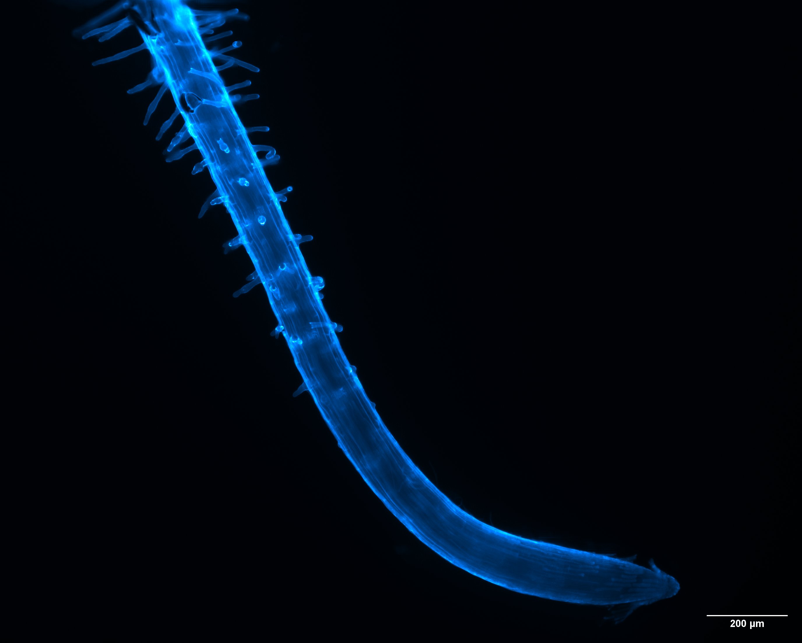
Locate the slender extensions in the genuine Arabidopsis fluorescence image. Compare the hair-bearing region with the comparatively hairless tip; the blue signal is not natural root colour.
Check the identification key
The slender projections along the root behind the tip are root hairs. The curved tip at the lower end is comparatively hairless. Each root hair is an epidermal-cell extension, not a separate multicellular lateral root. Blue fluorescence is an imaging signal, not natural root colour.
Look behind the tip
The supplied confocal image shows an Arabidopsis root curving towards a comparatively hairless tip. Slender lateral projections are visible farther behind that tip: these are root hairs. The image contrasts the hair-bearing region with the tip rather than implying hairs cover every root surface.
A root hair is an epidermal-cell extension. The complete root contains many cells, but that does not make each individual hair a multicellular root. Use the published specimen identity and the visible projections together.
Colour is an imaging signal
The blue appearance comes from fluorescence imaging. It does not show the natural daylight colour of the root or prove chlorophyll is present. The 200 μm scale bar gives a specimen distance, not a measured uptake rate.
The image does not resolve every membrane boundary or chemical transporter. Missing detail at this resolution does not mean a structure is absent. Identify the hairs first, then open the explanatory key to compare your reasoning.
Sources for this lesson
Pause and explain
Which visible region contains root hairs?
Worked example
Does the blue signal show that the root hair cells photosynthesise? Explain.
Show the worked solution
- Identify blue as a fluorescence imaging signal.
- Photosynthetic activity needs appropriate evidence, not this colour alone.
Answer No. Blue fluorescence does not establish chlorophyll or photosynthesis in these cells.
Try a topic test
Choose Easy, Normal or Difficult. Save your answers and open worked review after finishing.
Test this topic →From guided practice to a new situation
Write your answer and reasoning first. Use a hint only if you are stuck. The model solution is for self-checking; your written work is not automatically marked.
Identify the slender side projections and name their uptake materials.
Give me a hint
Use the image behind the comparatively hairless tip.
Compare my reasoning
- The side projections are root hairs.
- They support water and mineral-ion uptake.
Root hairs; they absorb water and mineral ions from soil solution.
Look for these in your work
- Locate projections
- State both materials
A hair appears half as long as the 200 μm bar in this image. Estimate its length and state a limitation.
Give me a hint
Use the bar only within this image and allow for projection.
Compare my reasoning
- Half of 200 μm is 100 μm.
- The image shows projected length; an out-of-plane or curved hair may be longer.
About 100 μm projected length; orientation/curvature limits the estimate of actual hair length.
Look for these in your work
- Calculate 100 μm
- State a relevant imaging limitation
Why is an image of a root tip alone weak evidence that a plant has no root hairs?
Give me a hint
Which root region normally bears the hairs?
Compare my reasoning
- Hairs are usually behind the growing tip.
- A tip-only field can omit that region.
The field may exclude the hair-bearing region, so it cannot establish absence throughout the plant.
Look for these in your work
- Locate the relevant region
- Avoid a whole-plant absence claim
Common mistakes
- Treating fluorescence colour as natural tissue colour.
- Assuming the hairless tip means the whole root lacks hairs.
What can you explain now?
Which visible region contains root hairs?
Compare with the explanation
The slender side projections behind the tip
Their location and specimen key identify them.
After trying it yourself, choose your next review. This is your self-assessment.
Your review choice appears on Today. Sign in to sync it across devices.
Make it stick
Describe how you distinguished a root hair from the root tip in the image.
If you needed the solution, close it and solve the problem again from a blank page. A correct answer is stronger when you can explain the reason for each step.