Identify the seven Core structures in a published eye image
Read all seven named targets in the unchanged source section.
Mapped to the named 0654 section using teaching from the corresponding single-science course. A 0654-specific review of every Core and Supplement objective is still in progress. Written practice is self-checked; it does not demonstrate supervised laboratory competence or award an official Cambridge grade.
Before you begin
- Recall receptors, electrical impulses, CNS organisation and the distinction between a stimulus and an effector response.
Work on paper. Try each question before revealing help, and explain why your method works.
Quick readiness check
Which printed label corresponds to the Core blind-spot region?
What you will learn
- Establish the source orientation
- Trace the seven required endpoints
Identify the seven Core structures in a published eye image
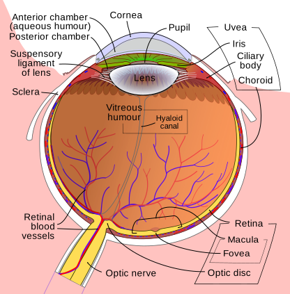
Published eye section: front top, posterior bottom. Seven Core targets include the anatomical blind-spot label Optic disc; Fovea is separate. Source image unchanged.
Enlarge image for inspection
Pan across the enlarged vector image to follow its leaders. Try identification before opening the key.
Check the identification key
Cornea: top curved cap. Iris: green tissue near front. Pupil: opening between iris edges, not its black leader marker. Lens: named body behind pupil. Retina: yellow inner posterior lining towards lower-right. Optic nerve: bundle leaving lower-left. Blind spot: printed Optic disc at the separate nerve exit.
Establish the source orientation
The unchanged published schematic by Rhcastilhos and Jmarchn is oriented with the front at the top and the posterior retina at the bottom. The Cornea leader identifies the curved front cap. Iris points to green tissue near the front, while Pupil points to the central opening between iris edges. The black endpoint circle is a leader marker, not a solid pupil structure.
Lens is named within the biconvex body immediately behind the pupil. Retina points to the yellow inner lining towards the posterior lower-right wall. Optic nerve identifies the yellow bundle leaving towards the lower-left. Follow each leader or printed in-structure label rather than naming a part from the nearest text alone.
Trace the seven required endpoints
The blind-spot region is printed as Optic disc in this source. Its leader ends at the retinal nerve-exit region near the leaving optic nerve. The Fovea leader ends separately within the macula; that extra Supplement target does not replace Optic disc in the seven Core identification list. Naming optic disc is an anatomical identification of the blind-spot region.
The source includes extra structures such as choroid, sclera and ciliary body. These do not all need to be named for this Core question, and they do not change the seven required targets. It is a published schematic rather than a specimen photograph; its colours and proportions are not measurements. Enlarge the vector source to trace the leaders and attempt identification before opening the separate key.
Sources for this lesson
Pause and explain
Which interpretation of Pupil and Iris is correct?
Worked example
Identify all seven required Core structures in the source image.
Show the worked solution
- Cornea, iris, pupil and lens are the front targets.
- Retina, optic nerve and Optic disc identify the lining, leaving nerve and blind-spot region.
Answer Cornea; iris; pupil; lens; retina; optic nerve; blind spot at the printed Optic disc.
Try a topic test
Choose Easy, Normal or Difficult. Save your answers and open worked review after finishing.
Test this topic →From guided practice to a new situation
Write your answer and reasoning first. Use a hint only if you are stuck. The model solution is for self-checking; your written work is not automatically marked.
Identify the seven required Core structures in the unchanged source.
Give me a hint
Include the source synonym for the blind-spot region.
Compare my reasoning
- Find cornea, iris, pupil and lens near the front.
- Find retina, optic nerve and Optic disc towards the posterior region.
Cornea; iris; pupil; lens; retina; optic nerve; blind spot at the printed Optic disc.
Look for these in your work
- Identify all seven
- Map Optic disc to blind spot
Locate the retinal lining and leaving optic nerve in this top-front source.
Give me a hint
Use endpoint position rather than nearest text.
Compare my reasoning
- Retina points to yellow inner lining towards lower-right posterior wall.
- Optic nerve is the bundle leaving towards lower-left.
Retina: yellow inner lining towards the lower-right posterior wall. Optic nerve: yellow bundle leaving towards the lower-left.
Look for these in your work
- Trace the retinal endpoint
- Identify the leaving nerve
Why does the separate Fovea label not supply the Core blind-spot identification?
Give me a hint
Compare receptor-rich specialised retina and nerve exit.
Compare my reasoning
- Fovea is a distinct cone-rich region.
- Optic disc is the receptor-free nerve-exit blind spot.
Fovea is a separate cone-rich retinal region; the Core blind-spot target is the printed Optic disc at the receptor-free nerve exit.
Look for these in your work
- Distinguish both locations
- Use the source blind-spot synonym
Common mistakes
- Naming the Fovea as the blind spot.
- Treating the black pupil leader marker as solid tissue.
What can you explain now?
Which interpretation of Pupil and Iris is correct?
Compare with the explanation
Pupil is the opening; Iris is the surrounding tissue
Follow the endpoints in the top/front of the image.
After trying it yourself, choose your next review. This is your self-assessment.
Your review choice appears on Today. Sign in to sync it across devices.
Make it stick
Identify all seven Core source targets including Optic disc as the blind-spot region.
If you needed the solution, close it and solve the problem again from a blank page. A correct answer is stronger when you can explain the reason for each step.