Identify vesicles gap and receptors in a published synapse image
Translate the unchanged source labels into the syllabus structures.
Mapped to the named 0654 section using teaching from the corresponding single-science course. A 0654-specific review of every Core and Supplement objective is still in progress. Written practice is self-checked; it does not demonstrate supervised laboratory competence or award an official Cambridge grade.
Before you begin
- Recall cell membranes, diffusion, sensitivity and the difference between cells, tissues and organs.
Work on paper. Try each question before revealing help, and explain why your method works.
Quick readiness check
What does Synaptic cleft name in this source?
What you will learn
- Read the enlarged axon-terminal view
- Separate the source illustration from a scale measurement
Identify vesicles gap and receptors in a published synapse image
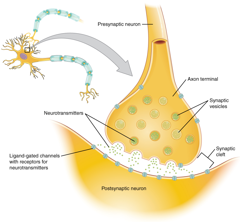
Unchanged OpenStax chemical-synapse illustration. Cleft means gap; receiving receptor-bearing channels are shown on the postsynaptic membrane. The inset arrow indicates enlargement.
Enlarge image for inspection
Scroll across the source to trace its leaders and gap bracket. Try identification before opening the key.
Check the identification key
Synaptic vesicles: round compartments within the upper terminal. Neurotransmitters: green particles in vesicles and cleft. Synaptic cleft: gap between membranes. Ligand-gated channels with receptors for neurotransmitters: turquoise receptor-bearing proteins on the lower postsynaptic membrane. The grey inset arrow marks enlargement, not an electrical impulse crossing the gap.
Read the enlarged axon-terminal view
The published OpenStax image shows an enlarged chemical synapse. The upper bulb is labelled Axon terminal and belongs to the presynaptic neurone. The lower receiving cell is labelled Postsynaptic neuron. The small whole-cell inset and grey magnification arrow locate the enlarged detail; that broad arrow is not an electrical impulse crossing the gap.
The printed Synaptic vesicles leaders end on round membrane-bounded compartments in the upper terminal. Neurotransmitters points to green particles inside a vesicle and in the space below. These are the molecules contained by the vesicles and released into the junction. The source shapes and colours make the container and contents distinguishable but do not establish their real relative size.
Separate the source illustration from a scale measurement
The printed Synaptic cleft bracket spans the space between the upper terminal and lower membrane. Cleft is the source name for the synaptic gap. The longer label Ligand-gated channels with receptors for neurotransmitters points to a turquoise protein shape in the postsynaptic membrane. These receptor-bearing proteins provide the receiving binding sites needed in the syllabus account.
The image illustrates a common receptor-bearing channel model; it does not establish that every receptor in all synapses must be this channel type. It also does not identify these two cells as sensory, relay or motor neurones. Read the actual labels and connected regions rather than inventing those roles from shape alone. Try the identification before opening the separate key, and enlarge the unchanged image to follow the small leaders.
Sources for this lesson
Pause and explain
Where does the receiving receptor-bearing channel leader end?
Worked example
Find vesicles, neurotransmitter, gap and receiving receptor proteins in the source image.
Show the worked solution
- Follow the vesicle and neurotransmitter leaders in the upper terminal.
- Read the cleft bracket and receptor-bearing channel leader on the lower membrane.
Answer Round upper compartments are vesicles; green particles are neurotransmitter; the cleft is the gap; the lower receptor-bearing channel proteins provide binding sites.
Try a topic test
Choose Easy, Normal or Difficult. Save your answers and open worked review after finishing.
Test this topic →From guided practice to a new situation
Write your answer and reasoning first. Use a hint only if you are stuck. The model solution is for self-checking; your written work is not automatically marked.
Identify the printed source labels that establish the required synaptic structures.
Give me a hint
Match the source wording to the syllabus names.
Compare my reasoning
- Synaptic vesicles contain the green neurotransmitter molecules.
- Synaptic cleft is the gap; receptor-bearing channels are on the postsynaptic membrane.
Synaptic vesicles containing neurotransmitters; Synaptic cleft for the gap; Ligand-gated channels with receptors for neurotransmitters for the receiving receptor proteins.
Look for these in your work
- Match all required features
- Keep the original source labels
Explain the difference between a round vesicle and a small green particle in the source view.
Give me a hint
Container and contained chemical are different.
Compare my reasoning
- The round membrane-bound structure is a vesicle.
- The green particle represents a neurotransmitter molecule.
The vesicle is a membrane-bounded container; the green particles represent neurotransmitter molecules stored inside or released from it.
Look for these in your work
- Separate container and contents
- Use the actual image key
Does the grey arrow from the whole-cell inset prove an electrical impulse jumps across the cleft?
Give me a hint
Read its purpose as enlargement of detail.
Compare my reasoning
- It connects the inset location with the magnified terminal view.
- Chemical transmission across the gap uses neurotransmitter molecules.
No. The grey arrow indicates magnification of a region; it is not evidence of an electrical impulse jumping across the cleft.
Look for these in your work
- Read magnification correctly
- Retain chemical gap transmission
Common mistakes
- Treating the grey enlargement arrow as gap-crossing electrical travel.
- Calling every synaptic receptor the same channel type.
What can you explain now?
Where does the receiving receptor-bearing channel leader end?
Compare with the explanation
On the postsynaptic membrane
That is the next cell’s receiving side.
After trying it yourself, choose your next review. This is your self-assessment.
Your review choice appears on Today. Sign in to sync it across devices.
Make it stick
Locate the required structures in the published image and explain the magnification arrow.
If you needed the solution, close it and solve the problem again from a blank page. A correct answer is stronger when you can explain the reason for each step.