Identify neurone roles in a published reflex image
Read the source leaders and connected pathway rather than guessing from cell shape.
Exam scope and assessment notes
Core and Extended · Biology 0970, 2026–2028. Use the stated specimen features and evidence. Diagrams are schematic. Written practice is self-checked; human subject-teacher review, supervised practical competence and cumulative assessment remain pending. Cambridge states that IGCSE 0970 has the same subject content as IGCSE 0610, with 9–1 grading. This StudyForA pathway maps the 2026–2028 syllabus only. This 9–1 graded qualification is available only in a limited number of administrative zones. Confirm that your centre can enter you for this code and exam series with Cambridge International. Cambridge has published a separate syllabus for 2029. StudyForA has not reconciled that version yet; this page supports exams in 2026, 2027 and 2028 only.
Before you begin
- Recall cell membranes, diffusion, sensitivity and the difference between cells, tissues and organs.
Work on paper. Try each question before revealing help, and explain why your method works.
Quick readiness check
What identifies the upper incoming path here?
What you will learn
- Trace the three source neurone labels
- Link image location with function
Identify neurone roles in a published reflex image
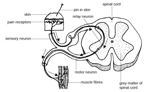
Unchanged published image: upper incoming sensory path, spinal grey-matter relay connector, lower outgoing motor path to muscle. The source neuron spelling is equivalent to neurone.
Enlarge image for inspection
Inspect the original 594-pixel source at its native size. Try identification before opening the key.
Check the identification key
Sensory neuron: upper incoming path from skin receptors towards cord. Relay neuron: short connector inside spinal grey matter. Motor neuron: lower outgoing cord-to-muscle path. Muscle fibres are the effector. Source neuron spelling is equivalent to neurone; the pin stimulus is for interpretation only.
Trace the three source neurone labels
The unchanged published illustration by Ruth Lawson, Otago Polytechnic, shows pain receptors in skin, incoming and outgoing nerve pathways, a spinal-cord section and muscle fibres. Its printed labels use neuron, the alternative spelling of neurone. The image is a simplified anatomical illustration rather than a specimen photograph or a microscopic view of individual cells.
The sensory neuron leader ends on the upper incoming path from the skin receptors towards the spinal cord. The relay neuron leader ends on the short connecting path within the grey matter of the cord. The motor neuron leader ends on the lower outgoing path from the cord towards the muscle fibres. Follow each leader to its endpoint before using its position on the page.
Link image location with function
The arrows and connections explain the functional identities: sensory carries receptor input towards CNS, relay links the central pathways, and motor carries output towards an effector. Here the muscle fibres are the effector. The source does not label cell bodies, myelin or dendrites in sufficient detail to identify every role by shape, so those additional features should not be invented from the picture.
The pin-in-skin stimulus is an interpretation example in the published image, not an instruction to perform a painful test. The pictured simple arc includes a relay neurone in the cord; it does not prove every reflex has that same circuit or that the brain can never receive information about a spinal reflex. Keep the source unchanged, enlarge it if a leader is hard to follow, and compare your role identifications with the separate key.
Pause and explain
Which source path is the motor neurone?
Worked example
Identify the three source-labelled neurone pathways.
Show the worked solution
- Upper incoming skin-to-cord path is sensory; short cord connector is relay.
- Lower outgoing cord-to-muscle path is motor.
Answer Sensory: upper incoming receptor-to-cord path; relay: short connector in spinal grey matter; motor: lower outgoing cord-to-muscle path.
Try a topic test
Choose Easy, Normal or Difficult. Save your answers and open worked review after finishing.
Test this topic →From guided practice to a new situation
Write your answer and reasoning first. Use a hint only if you are stuck. The model solution is for self-checking; your written work is not automatically marked.
Identify the sensory relay and motor pathways in the published image.
Give me a hint
Trace each printed leader and its connected route.
Compare my reasoning
- Upper incoming path reaches cord from skin receptors; short connector lies in cord grey matter.
- Lower outgoing path reaches muscle fibres.
Sensory is the upper receptor-to-cord route; relay is the short connector inside spinal grey matter; motor is the lower cord-to-muscle route.
Look for these in your work
- Identify all three roles
- Use leaders and functional connections
Explain why the lower outgoing path is motor rather than sensory in this source.
Give me a hint
Use its direction and endpoint.
Compare my reasoning
- It carries output from the cord.
- It leads towards the muscle-fibre effector.
It is the motor path because it leads from CNS towards the muscle-fibre effector, while the upper sensory path carries receptor input towards CNS.
Look for these in your work
- Use output direction
- Name the muscle effector
Can this picture establish that every reflex contains the same relay connection or that the pin stimulus should be copied in a learner experiment?
Give me a hint
A published illustration has a defined teaching scope.
Compare my reasoning
- It depicts one simplified supplied circuit.
- Its painful stimulus is for interpretation only.
No. It illustrates one supplied simple reflex circuit and is for image interpretation; it neither establishes every reflex circuit nor instructs learners to perform a painful stimulus test.
Look for these in your work
- Limit circuit generalisation
- Keep interpretation separate from performance
Common mistakes
- Naming roles only from unlabelled cell shape.
- Treating the source stimulus illustration as a practical instruction.
What can you explain now?
Which source path is the motor neurone?
Compare with the explanation
Lower outgoing cord-to-muscle path
The leader ends on the route towards the effector.
After trying it yourself, choose your next review. This is your self-assessment.
Your review choice appears on Today. Sign in to sync it across devices.
Make it stick
Identify the three source pathways and justify each from its functional connections.
If you needed the solution, close it and solve the problem again from a blank page. A correct answer is stronger when you can explain the reason for each step.