Identify valve categories in a prepared-heart photograph
Use verified numbered features in a historical anatomical photograph.
Exam scope and assessment notes
Extended only (Supplement) · Biology 0970, 2026–2028. Use the stated specimen features and evidence. Diagrams are schematic. Written practice is self-checked; human subject-teacher review, supervised practical competence and cumulative assessment remain pending. Cambridge states that IGCSE 0970 has the same subject content as IGCSE 0610, with 9–1 grading. This StudyForA pathway maps the 2026–2028 syllabus only. This 9–1 graded qualification is available only in a limited number of administrative zones. Confirm that your centre can enter you for this code and exam series with Cambridge International. Cambridge has published a separate syllabus for 2029. StudyForA has not reconciled that version yet; this page supports exams in 2026, 2027 and 2028 only.
Before you begin
- Recall valve positions in a mammalian-heart diagram.
Work on paper. Try each question before revealing help, and explain why your method works.
Quick readiness check
What are the two panels?
What you will learn
- Read the photographed specimen rather than the page layout
- Separate the valve from nearby landmarks
Read the photographed valves
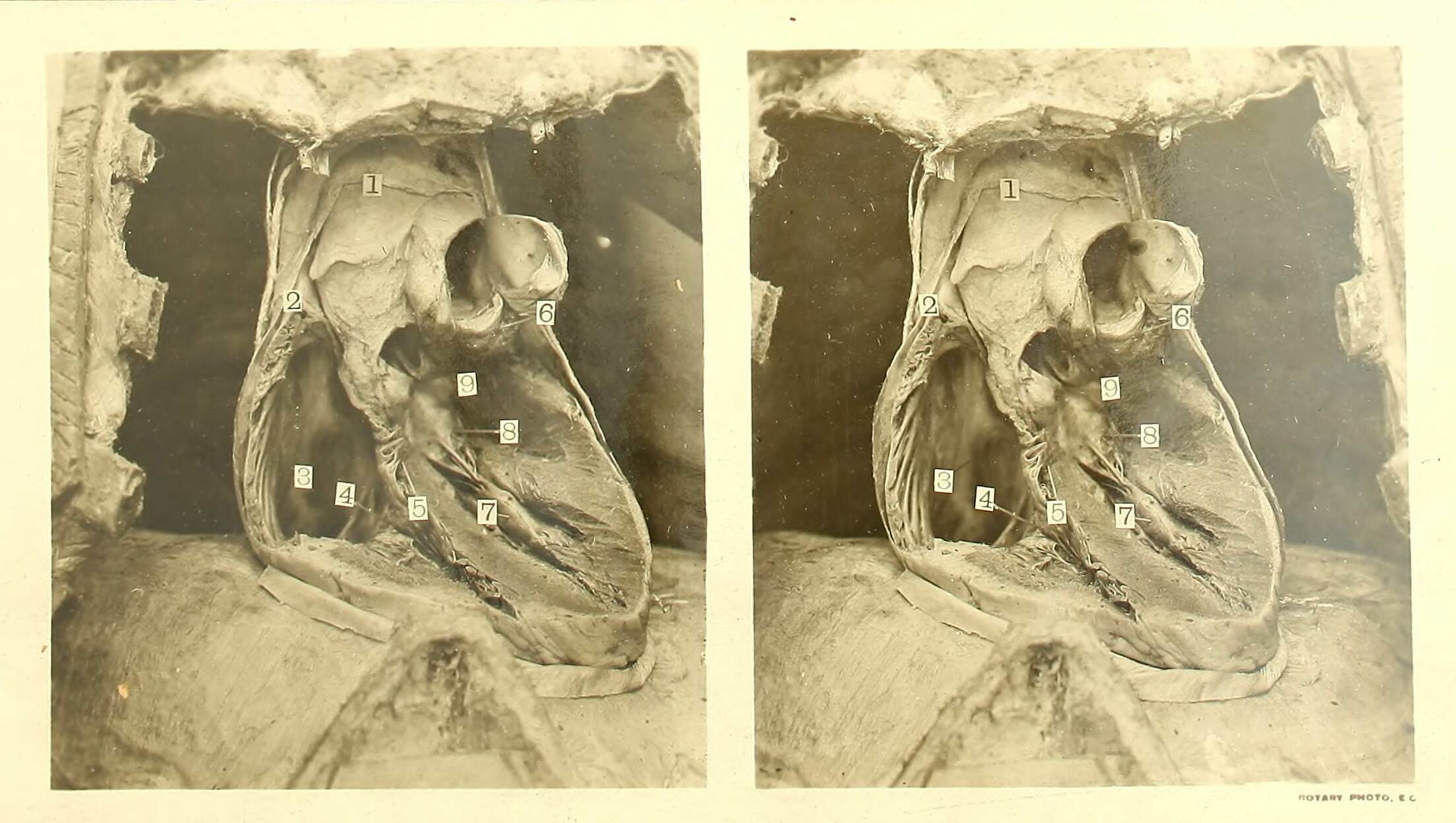
Waterston’s stereoscopic prepared-heart photograph: use verified features 4, 6 and 8 to identify atrioventricular and semilunar valves. Both panels show the same specimen.
Check the identification key
Both panels are viewpoints of the same prepared specimen. Source key: 4, septal cusp of tricuspid atrioventricular valve; 6, pulmonary semilunar valves; 8, aortic cusp of mitral atrioventricular valve; 5, interventricular septum. Label 9 lies below and to the right of the aortic valve; it is not a direct label of that valve. The mitral valve’s “aortic cusp” is still atrioventricular, not the aortic semilunar valve.
Read the photographed specimen rather than the page layout
The supplied image is a stereoscopic pair from David Waterston’s 1905 anatomical atlas. Both panels show the same prepared heart from slightly different viewpoints; they are not hearts from two animals. The specimen has been opened to expose internal structures, and the source provides a numbered identification key.
In that key, 4 identifies a cusp of the tricuspid atrioventricular valve, 6 identifies the pulmonary semilunar valves and 8 identifies a cusp of the mitral atrioventricular valve. Locate the numbered structures in the full-size image before opening the lesson’s key.
Separate the valve from nearby landmarks
A cusp is one of the valve’s flaps. Labels 4 and 8 therefore supply examples of the atrioventricular category, while 6 supplies the semilunar category. Category identification should use the source’s anatomical positions and key rather than the brightness or apparent size of a patch.
Label 5 identifies the interventricular septum. Label 9 lies below and to the right of the aortic valve: it is a neighbouring landmark, not a direct label of the aortic valve itself. Use the verified 4, 6 and 8 features for this exercise instead of inventing an identification for every numbered mark.
Pause and explain
Which verified feature is semilunar?
Worked example
Classify the source-key structures numbered 4, 6 and 8.
Show the worked solution
- 4 and 8 are tricuspid and mitral valve cusps at atrium–ventricle junctions.
- 6 identifies pulmonary semilunar valves at a ventricular exit.
Answer 4 and 8: atrioventricular; 6: semilunar.
Try a topic test
Choose Easy, Normal or Difficult. Save your answers and open worked review after finishing.
Test this topic →From guided practice to a new situation
Write your answer and reasoning first. Use a hint only if you are stuck. The model solution is for self-checking; your written work is not automatically marked.
Locate 4, 6 and 8 in the full-size source image, then classify them.
Give me a hint
Use the numbers and their verified source identities.
Compare my reasoning
- 4 and 8 identify atrioventricular cusps.
- 6 identifies pulmonary semilunar valves.
4 tricuspid atrioventricular; 6 pulmonary semilunar; 8 mitral atrioventricular.
Look for these in your work
- Locate the numbered structures
- Correctly classify both categories
Explain why 4 and 8 belong to one category despite looking different in the preparation.
Give me a hint
Their junction position defines the category.
Compare my reasoning
- They lie at right and left atrium–ventricle junctions.
- Differences in preparation or shape do not change that category.
Both are atrioventricular valve cusps at an atrium–ventricle junction, one on each side.
Look for these in your work
- Use anatomical position
- Do not classify by brightness or shape alone
A learner uses label 9 as a direct aortic-valve label. What does the source key actually support?
Give me a hint
Read the exact landmark relationship.
Compare my reasoning
- 9 lies below and to the right of the aortic valve.
- A neighbouring landmark is not a direct valve label.
9 marks a position below/right of the aortic valve, not the valve itself; use the verified 6 feature for semilunar identification in this exercise.
Look for these in your work
- State the relationship accurately
- Use a verified valve feature
Common mistakes
- Treating the two stereo panels as different animals.
- Calling label 9 the aortic valve itself.
What can you explain now?
Which verified feature is semilunar?
Compare with the explanation
6: pulmonary semilunar valves
The source identifies the pulmonary exit valves.
After trying it yourself, choose your next review. This is your self-assessment.
Your review choice appears on Today. Sign in to sync it across devices.
Make it stick
Locate 4, 6 and 8 again with the key closed and explain their categories.
If you needed the solution, close it and solve the problem again from a blank page. A correct answer is stronger when you can explain the reason for each step.