Identify transport tissues in a sunflower stem micrograph
Distinguish conducting xylem and phloem from surrounding support tissues in a real bundle.
Exam scope and assessment notes
Core and Extended · Biology 0970, 2026–2028. Use the stated specimen features and evidence. Diagrams are schematic. Written practice is self-checked; human subject-teacher review, supervised practical competence and cumulative assessment remain pending. Cambridge states that IGCSE 0970 has the same subject content as IGCSE 0610, with 9–1 grading. This StudyForA pathway maps the 2026–2028 syllabus only. This 9–1 graded qualification is available only in a limited number of administrative zones. Confirm that your centre can enter you for this code and exam series with Cambridge International. Cambridge has published a separate syllabus for 2029. StudyForA has not reconciled that version yet; this page supports exams in 2026, 2027 and 2028 only.
Before you begin
- Complete the stem diagram lesson.
Work on paper. Try each question before revealing help, and explain why your method works.
Quick readiness check
What is the usual xylem cargo?
What you will learn
- Locate the thick-walled xylem openings
- Distinguish phloem from its fibre cap
Sunflower stem: one vascular bundle
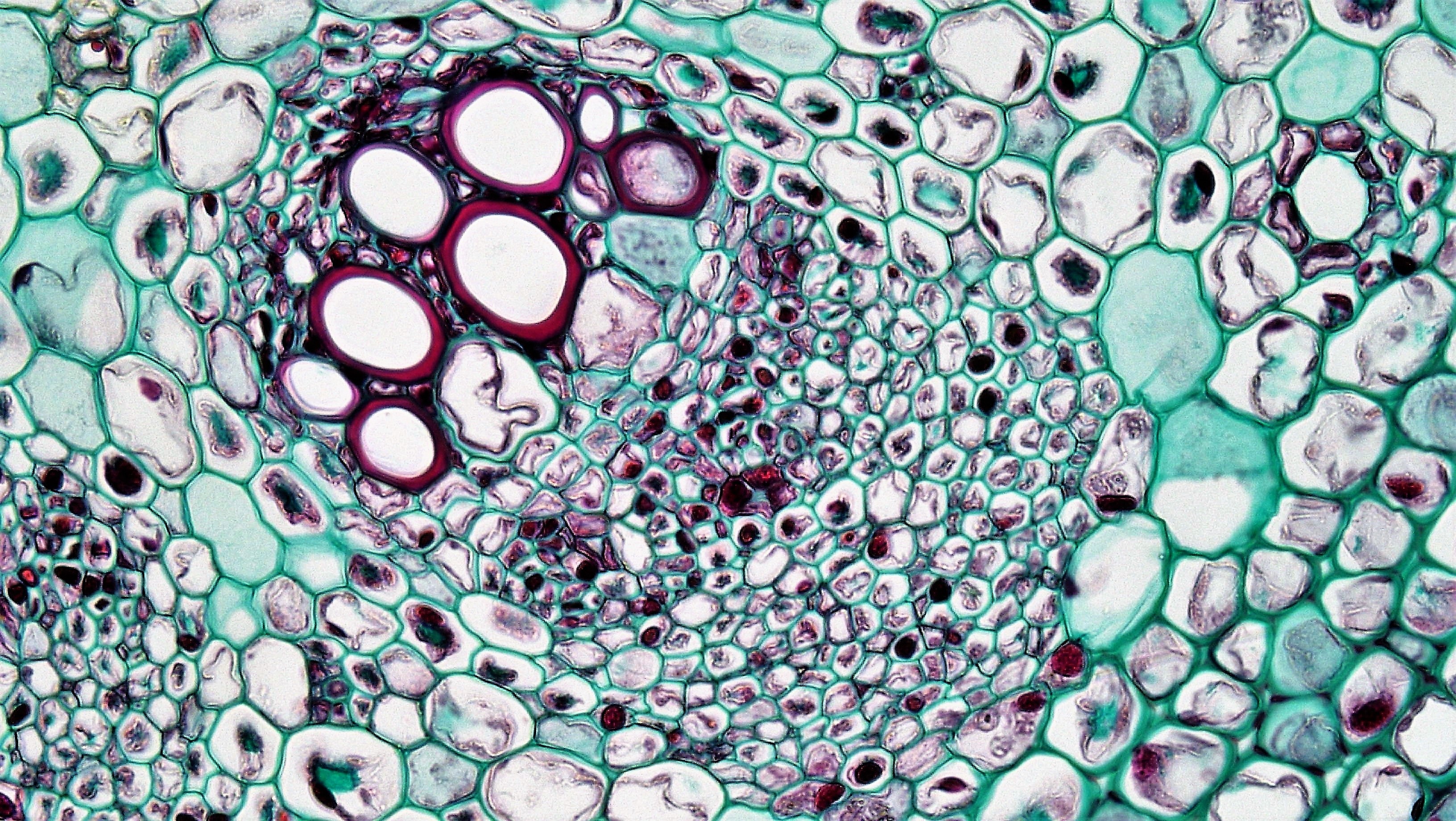
Use vessel structure and the source description to locate xylem and conducting phloem. The supporting fibre cap is distinct from the conducting phloem layer.
Check the identification key
The large thick-walled vessel openings identify inner xylem. Conducting phloem lies outside this region. In the source description, the outside-to-inside order is supporting fibre cap, conducting phloem, thin cambium, xylem. The cap is distinct from conducting phloem; this close-up does not show the whole bundle ring.
Read the section as a bundle
This Helianthus (sunflower) preparation shows a primary-growth bundle in an older herbaceous stem. The conspicuous large, thick red-walled vessel openings identify its inner xylem region. The conducting phloem layer lies outside the xylem, with a narrow intervening cambium region in the source description.
The outside of the bundle also has a supporting fibre cap. These fibres and the conducting phloem are not interchangeable structures. Find the smaller conducting cells just outside xylem rather than assigning every thick-walled outer cell to sucrose transport.
Match anatomy to the task
The source describes this stem as herbaceous and without the secondary wood arrangement of a woody stem. This makes it an appropriate non-woody dicot example; “older stem” does not automatically mean a tree’s growth rings.
Use inside/outside relationships rather than a fixed top-of-page rule. The frame is a close-up, and the complete ring of bundles is not shown. A real specimen can support bundle identification without containing the whole organ in the image.
Pause and explain
Which feature supports xylem identification here?
Worked example
A learner labels the outer support cap “the sucrose-conducting phloem”. What should be changed?
Show the worked solution
- Separate the supporting fibres from the deeper conducting phloem.
- Use the source-described tissue layer outside xylem for transport.
Answer The support cap is not the conducting layer. Sucrose moves in the phloem layer outside xylem, beneath the outer fibre cap.
Try a topic test
Choose Easy, Normal or Difficult. Save your answers and open worked review after finishing.
Test this topic →From guided practice to a new situation
Write your answer and reasoning first. Use a hint only if you are stuck. The model solution is for self-checking; your written work is not automatically marked.
Name the tissue containing the conspicuous red-walled vessel openings and its transport cargo.
Give me a hint
Combine anatomical evidence with function.
Compare my reasoning
- The vessel openings belong to xylem.
- Xylem carries water/mineral ions.
Xylem; water and dissolved mineral ions.
Look for these in your work
- Name the tissue
- State both types of cargo
State the relative order of fibre cap, conducting phloem and xylem from outside to inside.
Give me a hint
The fibre cap is outermost of these three.
Compare my reasoning
- Begin with the outer support cap.
- Next place conducting phloem, then inner xylem.
Fibre cap → conducting phloem → xylem, omitting the thin intervening cambium from this three-item list.
Look for these in your work
- Keep support and conducting regions distinct
- Give the correct order
Does this close-up demonstrate that the stem has only one vascular bundle? Explain.
Give me a hint
What does the image frame exclude?
Compare my reasoning
- The image isolates one bundle region.
- The whole cross-section can contain other bundles outside the frame.
No. One bundle in a close-up does not mean one bundle in the entire stem.
Look for these in your work
- State the field-of-view limit
- Avoid an organ-wide inference
Common mistakes
- Confusing phloem fibres with conducting phloem.
- Inferring growth rings merely from the word older.
What can you explain now?
Which feature supports xylem identification here?
Compare with the explanation
Large thick-walled vessel openings in the inner bundle region
Use structure, position and the published key together.
After trying it yourself, choose your next review. This is your self-assessment.
Your review choice appears on Today. Sign in to sync it across devices.
Make it stick
Explain why a phloem fibre cap is not the same identification as conducting phloem.
If you needed the solution, close it and solve the problem again from a blank page. A correct answer is stronger when you can explain the reason for each step.