Identify ovary and ovules in a real cut-section photograph
Locate enclosed structures without confusing a flower stalk with a style.
Exam scope and assessment notes
Core and Extended · Biology 0970, 2026–2028. Use the stated specimen features and evidence. Diagrams are schematic. Written practice is self-checked; human subject-teacher review, supervised practical competence and cumulative assessment remain pending. Cambridge states that IGCSE 0970 has the same subject content as IGCSE 0610, with 9–1 grading. This StudyForA pathway maps the 2026–2028 syllabus only. This 9–1 graded qualification is available only in a limited number of administrative zones. Confirm that your centre can enter you for this code and exam series with Cambridge International. Cambridge has published a separate syllabus for 2029. StudyForA has not reconciled that version yet; this page supports exams in 2026, 2027 and 2028 only.
Before you begin
- Recall gametes, nuclear fusion, cellular respiration and the distinction between reproduction and growth.
Work on paper. Try each question before revealing help, and explain why your method works.
Quick readiness check
Which supported feature lies inside the cut-open ovary?
What you will learn
- Find the ovary wall around the central cavity
- Distinguish visible anatomy from reproductive events
Identify ovary and ovules in a real cut-section photograph
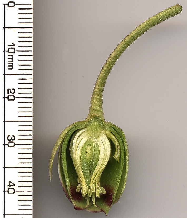
Helleborus foetidus cut section: green ovary wall encloses three visible pale ovules. The upward stalk is the flower stalk, not a style. No nuclear fusion is resolved. Original teaching data.
Ovary and ovules · Helleborus foetidus

Enlarge image for inspection
Inspect the unchanged original at its native pixel size. Scroll inside the image to follow the structures.
Check the English identification key
- ovary: Central cut-open green organ; its wall encloses the cavity containing the pale ovules.
- ovary wall: Green tissue on either side of the central cavity.
- ovules: Three pale round bodies visible within the central cavity.
The pale bodies are identified as ovules; nuclei, fertilisation and mature seeds are not resolved. Three ovules are visible in this section; this is not the total for every flower or ovary. The long upward stalk is the flower stalk, not a style. Use this photograph for ovary and ovule identification; its outer flower parts differ from the simplified cutaway.
| Feature | Meaning |
|---|---|
| Ovary wall | Green tissue enclosing the central cavity |
| Ovules | Three pale bodies visible inside the cavity |
| Orientation | Upward long stalk is the flower stalk |
| Limit | No observed nuclei, fusion or mature seeds |
Find the ovary wall around the central cavity
The published section photograph is identified by its original source as Helleborus foetidus. Focus on the cut-open green reproductive structure near the lower centre of the displayed image. Green tissue bounds a central cavity, and three pale round bodies are visible within that cavity. The documented identification key locates the enclosing ovary wall and these ovules, complementing the external dissection that hides internal structures.
An ovary is the enclosing organ; an ovule is one of the structures inside it. The ovule contains the female reproductive cell whose nucleus can take part in fertilisation. The photograph supports identifying the visible ovules, but does not resolve their individual cells or nuclei. A rounded outline in a different region is not sufficient evidence for another ovule without a supported anatomical relationship.
Distinguish visible anatomy from reproductive events
Orientation is especially important in this source. The long green stalk extending toward the top of the displayed photograph is the flower stalk, not the style of an upright classroom cutaway. Do not transfer top-to-bottom positions from the schematic into a section turned another way. Use the cavity, its surrounding wall and the verified key rather than a universal rule that the highest thin structure must be the style.
Three visible ovules are a count within this photographed view, not a claim about the total in every ovary or every flower. The section does not establish successful nuclear fusion, an embryo or a mature seed. Enlargement can help distinguish the pale bodies from the green wall but cannot add microscopic events that the original did not record. The learning target here is real-image identification and the ovary–ovule relationship.
Pause and explain
Does this photograph show fertilisation occurring?
Worked example
The section contains three keyed pale bodies enclosed by a green wall. Name the bodies and enclosing organ.
Show the worked solution
- The enclosed bodies are ovules.
- The enclosing organ is an ovary.
Answer Three visible ovules within the cut-open ovary.
Try a topic test
Choose Easy, Normal or Difficult. Save your answers and open worked review after finishing.
Test this topic →From guided practice to a new situation
Write your answer and reasoning first. Use a hint only if you are stuck. The model solution is for self-checking; your written work is not automatically marked.
Describe how to distinguish the ovary wall from the ovules in the photograph.
Give me a hint
Use enclosure and the verified internal key.
Compare my reasoning
- Green tissue bounds the central cavity.
- Three pale round bodies lie inside the cavity.
The surrounding green tissue is ovary wall; the three pale bodies enclosed within the cavity are visible ovules.
Look for these in your work
- Locate the enclosure
- Name the internal bodies
Why is the long upward green stalk not labelled style in this image?
Give me a hint
Check the source orientation rather than the schematic position.
Compare my reasoning
- The source identifies it as the flower stalk.
- The section is oriented differently from an upright flower diagram.
It is the flower stalk. A structure being high and slender does not override its source-key identity or make it a style.
Look for these in your work
- Use the documented identity
- Reject position-only identification
A learner writes “three mature seeds show three completed fertilisations.” Correct the statement.
Give me a hint
Name visible structures before inferring events.
Compare my reasoning
- The visible bodies are keyed as ovules.
- Fusion, embryo development and maturity are not shown.
The image shows three visible ovules within an ovary. It does not establish completed fertilisation, embryos or mature seeds.
Look for these in your work
- Correct the anatomical name
- Keep reproductive events unproven
Common mistakes
- Calling the upward flower stalk a style.
- Calling the ovules mature seeds or proven embryos.
What can you explain now?
Does this photograph show fertilisation occurring?
Compare with the explanation
No; nuclear fusion is not resolved
Visible organs do not establish the cellular event.
After trying it yourself, choose your next review. This is your self-assessment.
Your review choice appears on Today. Sign in to sync it across devices.
Make it stick
Identify ovary and ovules from enclosure in a section, rather than schematic orientation.
If you needed the solution, close it and solve the problem again from a blank page. A correct answer is stronger when you can explain the reason for each step.