Locate the fovea in a published eye image and explain its function
Follow the Fovea endpoint within the macula rather than the Optic disc endpoint.
Exam scope and assessment notes
Extended only (Supplement) · Biology 0970, 2026–2028. Use the stated specimen features and evidence. Diagrams are schematic. Written practice is self-checked; human subject-teacher review, supervised practical competence and cumulative assessment remain pending. Cambridge states that IGCSE 0970 has the same subject content as IGCSE 0610, with 9–1 grading. This StudyForA pathway maps the 2026–2028 syllabus only. This 9–1 graded qualification is available only in a limited number of administrative zones. Confirm that your centre can enter you for this code and exam series with Cambridge International. Cambridge has published a separate syllabus for 2029. StudyForA has not reconciled that version yet; this page supports exams in 2026, 2027 and 2028 only.
Before you begin
- Recall receptors, electrical impulses, CNS organisation and the distinction between a stimulus and an effector response.
Work on paper. Try each question before revealing help, and explain why your method works.
Quick readiness check
Which printed source label identifies the blind-spot region rather than fovea?
What you will learn
- Read the actual published source orientation
- Link the identified fovea with cone-rich vision
Locate the fovea in a published eye image and explain its function
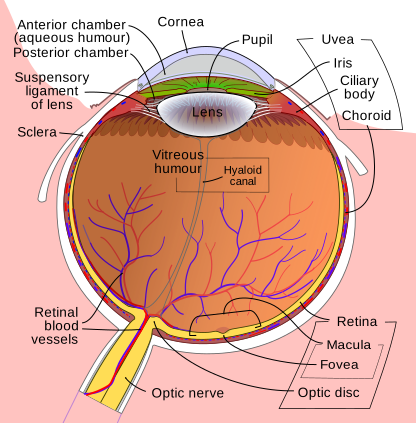
Published section: front top, retina bottom. Follow Fovea to its pit within Macula; Optic disc is a separate nerve exit. The image does not directly show cone density.
Enlarge image for inspection
Pan across the enlarged vector image to follow its leaders. Try identification before opening the key.
Check the identification key
Fovea: named leader to a small retinal pit towards lower central/right within Macula. Macula is the broader region. Optic disc: separate lower-left nerve exit and receptor-free blind spot. Dense cones support detailed central colour vision, but individual cones are not drawn in this gross schematic.
Read the actual published source orientation
The unchanged published eye section by Rhcastilhos and Jmarchn has the front at the top and the posterior retina towards the bottom. This differs from the left-front original diagram. Use the Cornea and Lens labels to establish orientation before following the retinal leaders; page direction alone is not an anatomical identity.
The printed Fovea leader ends at a small retinal pit towards the lower central/right region, within the broader area labelled Macula. The Optic disc leader ends separately towards the lower-left nerve-exit region. Identify the Fovea endpoint itself rather than the entire Macula bracket or the nearest text beside the optic nerve.
Link the identified fovea with cone-rich vision
The fovea is cone-rich and supports detailed, sharp central colour vision. The optic disc is the receptor-free blind spot. The source image identifies positions but does not directly depict individual cones or their density; those functional and receptor explanations come from the accompanying teaching rather than a claim that cells can be counted in the picture.
Black circles mark leader endpoints, not literal holes or receptor cells. The published colours and tissue thicknesses are schematic and not to scale. Enlarging the unchanged source helps trace the Fovea and Optic disc leaders, while the separate identification key can remain closed until an attempted answer. Extra source labels exceed the required identification scope.
Pause and explain
Which source-reading method identifies the fovea correctly?
Worked example
Locate the published Fovea and give its function.
Show the worked solution
- Follow the Fovea leader into the macular retinal pit, separate from Optic disc.
- State dense cones and detailed central colour vision.
Answer Fovea: distinct retinal pit within macula; cone-rich for detailed central colour vision.
Try a topic test
Choose Easy, Normal or Difficult. Save your answers and open worked review after finishing.
Test this topic →From guided practice to a new situation
Write your answer and reasoning first. Use a hint only if you are stuck. The model solution is for self-checking; your written work is not automatically marked.
Locate the fovea in the published image and give its function.
Give me a hint
Follow the named leader rather than its text position.
Compare my reasoning
- Fovea ends at the small pit within macula towards lower central/right retina.
- Dense cones support detailed central colour vision.
The Fovea leader identifies the retinal pit within the macula, separate from Optic disc; the cone-rich fovea supports detailed central colour vision.
Look for these in your work
- Trace the actual endpoint
- Explain the dense-cone function
Distinguish the printed Macula and Fovea targets.
Give me a hint
One identifies a broader retinal region.
Compare my reasoning
- Macula labels the surrounding broader area.
- Fovea identifies the smaller specialised retinal pit within it.
Macula is the broader region; the Fovea leader identifies the smaller retinal pit within it. They are not interchangeable exact identification targets.
Look for these in your work
- Distinguish broader region and target
- Follow the Fovea leader
Can individual cone density be counted from this published gross eye section? Explain how the function answer is supported instead.
Give me a hint
Separate visible anatomical position from accompanying receptor teaching.
Compare my reasoning
- The image identifies gross locations, not individual photoreceptors.
- The teaching explains dense cones and central colour vision.
No. This gross schematic does not show countable cones; the dense-cone function is supplied by the accompanying receptor teaching, while the image identifies location.
Look for these in your work
- State image resolution and scope
- Separate location evidence from function teaching
Common mistakes
- Using the Macula bracket as the exact Fovea endpoint.
- Inferring visible receptor counts from this gross eye section.
What can you explain now?
Which source-reading method identifies the fovea correctly?
Compare with the explanation
Trace the Fovea leader to its retinal pit within the macula
Its endpoint is separate from the optic disc.
After trying it yourself, choose your next review. This is your self-assessment.
Your review choice appears on Today. Sign in to sync it across devices.
Make it stick
Trace the published Fovea endpoint and distinguish its function from the optic-disc blind spot.
If you needed the solution, close it and solve the problem again from a blank page. A correct answer is stronger when you can explain the reason for each step.