Identify bronchioles alveoli and capillaries in a published image
Follow the air branches and the distinct blood network in the magnified respiratory-zone illustration.
Core and Extended; Biology 0610, 2026–2028. Use the stated specimen features and evidence. Diagrams are schematic. Written practice is self-checked
Before you begin
- Recall alveolar and capillary compartments.
Work on paper. Try each question before revealing help, and explain why your method works.
Quick readiness check
Which structure normally contains air?
What you will learn
- Locate the exchange region
- Keep air and blood routes distinct
Identify bronchioles alveoli and capillaries in a published image
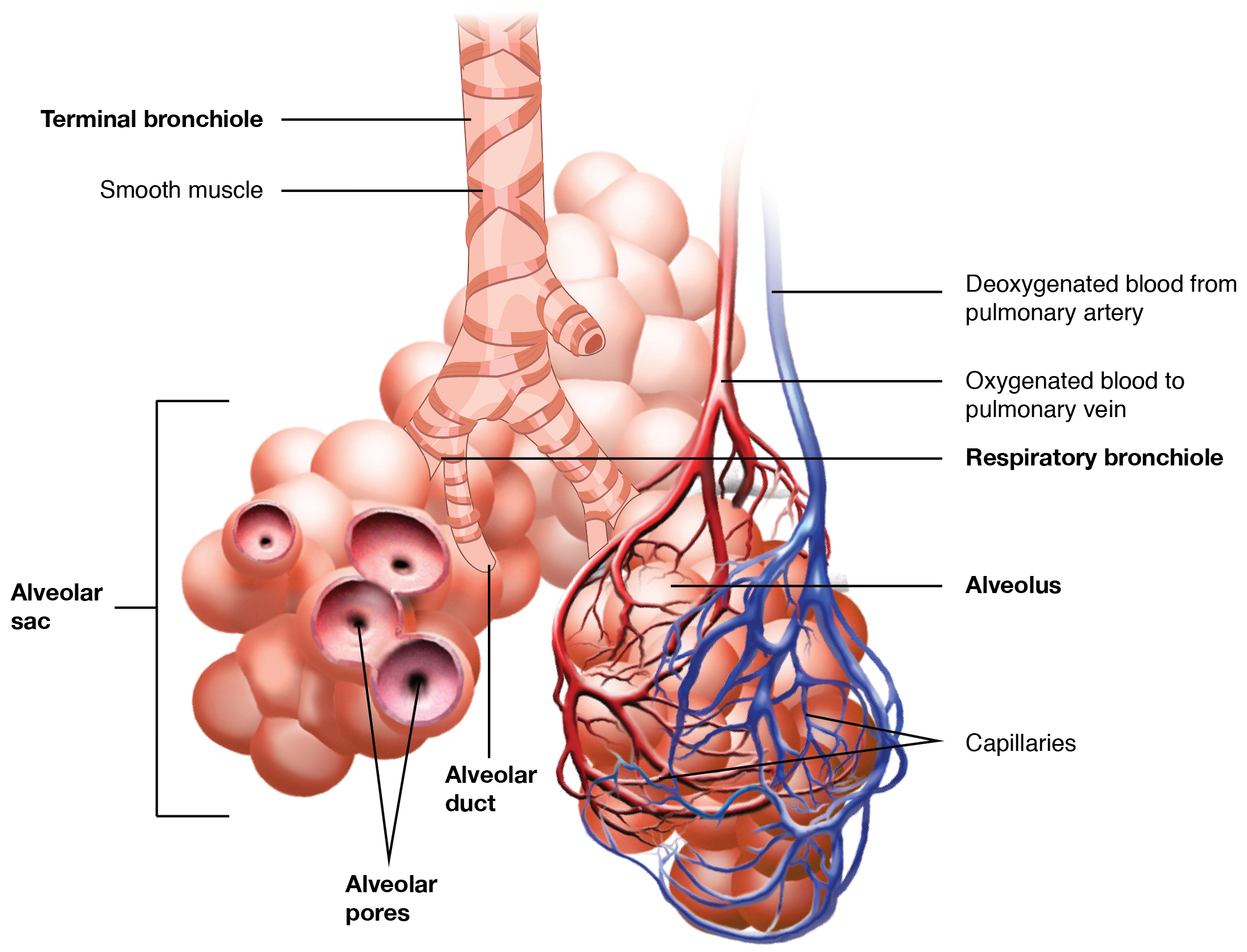
The published magnified view shows small air branches, alveolar spaces and the surrounding capillary blood network. Extra source labels are beyond this task.
Enlarge image for inspection
Scroll across the whole source image to inspect its labels. Try identification before opening the key.
Check the identification key
bronchioles: Small pink branching tubes running from the upper Terminal bronchiole to the labelled Respiratory bronchiole. alveoli: Rounded pink sacs at the tube ends. Alveolus points to one sac; Alveolar sac brackets a cluster on image left. capillaries: Fine red-and-blue branching network wrapped tightly around the sacs on image right, indicated by Capillaries. The two larger vessels are also labelled by blood direction. A magnified anatomical illustration, not a photograph. Extra labels (alveolar duct, pores and smooth muscle) exceed the requested identification list. Colours distinguish blood oxygenation; they are a diagram convention.
Locate the exchange region
The Respiratory Zone illustration shows terminal and respiratory bronchioles leading towards clusters of alveoli. The rounded pink air spaces at the ends are alveoli; the fine network surrounding the sacs on the right is labelled capillaries. Use the enlargement to follow each label to its structure rather than treating all branches as one type of tube.
An alveolus is one air sac; the image’s alveolar-sac bracket groups several. Extra labels such as ducts, pores and smooth muscle extend beyond this identification task. The published image helps recognise required structures in a different representation, while the original exchange diagram makes the two compartments easier to separate.
Keep air and blood routes distinct
The small air branches continue into the alveolar spaces. The capillary network lies around those spaces and connects to the labelled blood vessels. Oxygen and carbon dioxide exchange across their close boundaries; bulk air does not travel down the red and blue vessel network as though it were a bronchiole.
Red and blue distinguish the blood-route convention in this illustration; real blood is not blue. The view is enlarged without a stated anatomical scale and is not a clinical specimen. It demonstrates arrangement rather than diagnosing a lung, counting every alveolus or determining capillary diameter from the number of pixels.
Pause and explain
Which relationship matches this image?
Worked example
Identify the fine network around the alveoli on image right.
Show the worked solution
- Find the Capillaries label.
- Follow its pointer to the small surrounding blood routes.
Answer Associated capillaries.
Try a topic test
Choose Easy, Normal or Difficult. Save your answers and open worked review after finishing.
Test this topic →From guided practice to a new situation
Write your answer and reasoning first. Use a hint only if you are stuck. The model solution is for self-checking; your written work is not automatically marked.
Identify a bronchiole, an alveolus and associated capillaries in the source image.
Give me a hint
Trace labels in the enlargement.
Compare my reasoning
- Small pink branching tubes are bronchioles.
- Rounded sacs are alveoli; their surrounding fine blood network is capillaries.
Bronchiole: small air branch; alveolus: rounded air sac; capillaries: fine surrounding blood network.
Look for these in your work
- Identify all three
- Separate air and blood
Compare the air route and blood route at one alveolus.
Give me a hint
Use their connections.
Compare my reasoning
- Air arrives through a bronchiole into the alveolus.
- Blood passes through the surrounding capillary network.
Air reaches the alveolus through a bronchiole; blood flows in its associated capillaries across a separating exchange boundary.
Look for these in your work
- Describe both routes
- Retain the boundary
A learner identifies a large blue vessel as a bronchiole because it branches. What stronger evidence corrects the answer?
Give me a hint
Branching alone is shared by several routes.
Compare my reasoning
- The source labels and continuity place it in the blood route.
- Bronchioles connect to the air spaces.
Use the labelled blood connection to identify a vessel; branching and colour alone do not identify a bronchiole.
Look for these in your work
- Use route evidence
- Reject shape alone
Common mistakes
- Calling every small branch a bronchiole.
- Treating blue as the real colour of blood.
What can you explain now?
Which relationship matches this image?
Compare with the explanation
Bronchioles lead to alveoli; capillaries surround them
Air and blood routes are distinct.
After trying it yourself, choose your next review. This is your self-assessment.
Your review choice appears on Today. Sign in to sync it across devices.
Make it stick
Find the three structures and explain which route carries air.
If you needed the solution, close it and solve the problem again from a blank page. A correct answer is stronger when you can explain the reason for each step.