Identify transport tissues in a buttercup root micrograph
Use a genuine herbaceous dicot root image and its published specimen description.
Core and Extended; Biology 0610, 2026–2028. Use the stated specimen features and evidence. Diagrams are schematic. Written practice is self-checked
Before you begin
- Complete the dicot root diagram lesson.
Work on paper. Try each question before revealing help, and explain why your method works.
Quick readiness check
What is a lumen?
What you will learn
- Identify visible xylem vessel openings
- Use specimen evidence to locate phloem
Buttercup root: real tissue image
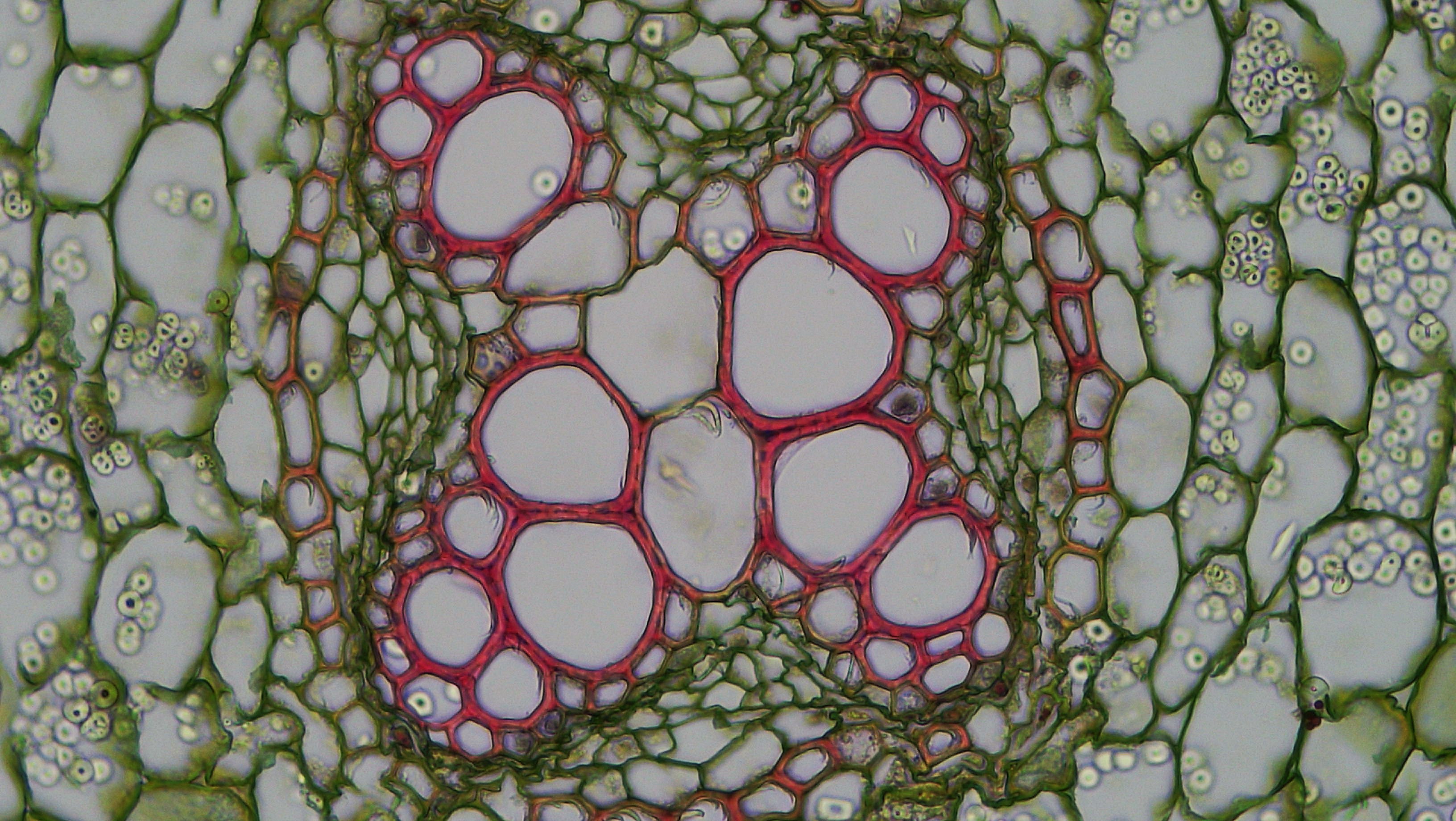
Identify the central xylem region and phloem patches before checking the key. This is a close view of a herbaceous dicot root, not the whole organ.
Check the identification key
The large red-walled openings form the central xylem region. The source identifies green-stained phloem groups between the xylem arms. Other surrounding cells also stain green, so colour alone is not sufficient.
A real section is less regular than a model
The supplied Ranunculus (buttercup) root image is a close view of the central transport region. Large openings with conspicuous red-stained walls belong to the xylem region. Smaller green-stained groups between its arms include phloem, as identified by the specimen source.
Some root tissues are outside the frame. The absence of a whole epidermal outline in this close-up does not mean that the original root lacked an epidermis. Use the source description together with visible position and structure.
Evidence has limits
Compare the image with the schematic: irregular outlines replace the neat star, but xylem and phloem remain distinct tissues. Stain colours apply to this preparation only. Many other cells also appear green, so green colour alone does not identify phloem.
A still section shows anatomy. It does not show moving water or measure transport rate. Begin with your own tissue identification, then open the identification key to compare the source-based interpretation.
Pause and explain
Which identification uses the strongest supplied evidence?
Worked example
Why are the large red-walled openings evidence for xylem in this image, but not a transport-rate measurement?
Show the worked solution
- Their position and thick-walled vessel openings match the published xylem identification.
- A still photograph records structure, not volume moved per time.
Answer They support xylem identification; they do not measure water-flow rate.
Try a topic test
Choose Easy, Normal or Difficult. Save your answers and open worked review after finishing.
Test this topic →From guided practice to a new situation
Write your answer and reasoning first. Use a hint only if you are stuck. The model solution is for self-checking; your written work is not automatically marked.
Locate a large red-walled opening near the central region. State the tissue and one transported material.
Give me a hint
Use the source key for this preparation.
Compare my reasoning
- Identify the opening as part of xylem.
- Give water or a dissolved mineral ion as cargo.
Xylem; it transports water and dissolved mineral ions.
Look for these in your work
- Identify xylem
- Give suitable cargo
Where should you look for phloem in this root section, and why is “all green areas” inadequate?
Give me a hint
Compare the spaces between xylem arms with the wider green surroundings.
Compare my reasoning
- The source identifies phloem between xylem arms.
- Cortex and other cells can also be green.
Look between the central xylem arms. Green stain alone is not sufficient to assign every green area to phloem.
Look for these in your work
- Locate inter-arm tissue
- Reject a colour-only rule
A close-up has no root hairs in its frame. Can you conclude that the plant has no root hair cells? Explain.
Give me a hint
Distinguish the field of view from the whole root.
Compare my reasoning
- This field shows internal central tissues.
- Root hairs occur on the external epidermis in the appropriate root region.
No. The central close-up does not include the relevant outer surface; missing from the frame is not absent from the plant.
Look for these in your work
- Use the field-of-view limit
- Locate root hairs externally
Common mistakes
- Calling every green cell phloem.
- Treating visible lumen area as a measured flow rate.
What can you explain now?
Which identification uses the strongest supplied evidence?
Compare with the explanation
Position and vessel structure plus the specimen key
The image and source support the tissue assignment together.
After trying it yourself, choose your next review. This is your self-assessment.
Your review choice appears on Today. Sign in to sync it across devices.
Make it stick
Describe two pieces of evidence for xylem in the buttercup image, and one limit of a still image.
If you needed the solution, close it and solve the problem again from a blank page. A correct answer is stronger when you can explain the reason for each step.