Recognise coronary arteries on the heart surface
Connect a genuine mammalian-heart photograph with the supply of the heart muscle itself.
Core and Extended; Biology 0610, 2026–2028. Use the stated specimen features and evidence. Diagrams are schematic. Written practice is self-checked
Before you begin
- Recall the muscular wall and the need for cellular respiration.
Work on paper. Try each question before revealing help, and explain why your method works.
Quick readiness check
What tissue receives the coronary artery supply?
What you will learn
- The pump muscle needs its own blood supply
- State what this photograph can and cannot show
Coronary vessels: an external view
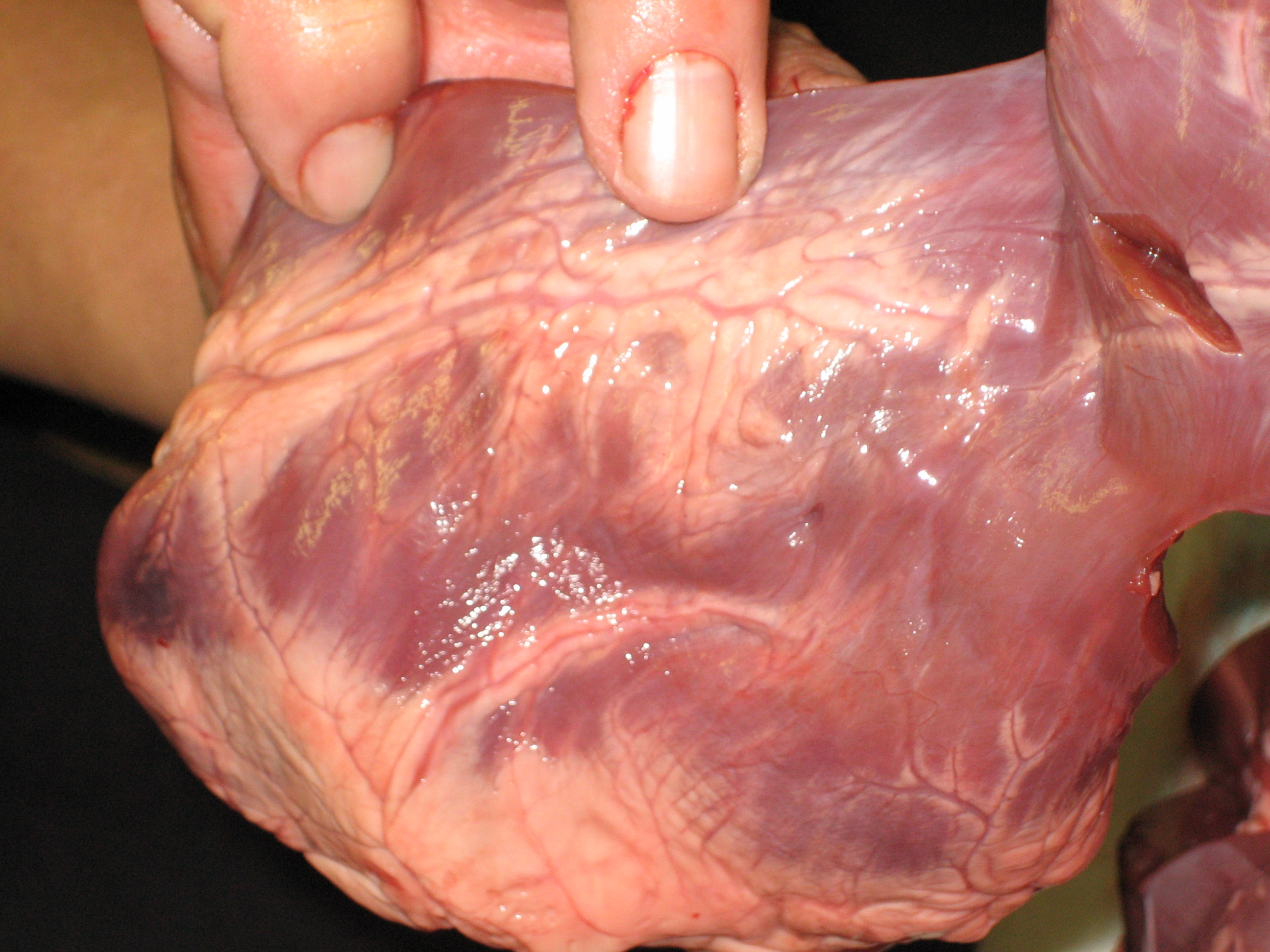
Prepared pig-heart photograph. Recognise the branching coronary supply on the heart surface; this view does not demonstrate internal chambers or disease.
Check the identification key
The credited source identifies coronary arteries. Recognise the exposed branching surface supply, including the prominent vessel in a surface groove. Do not label every pale line as an artery or name unverified individual branches. The vessels supply the heart muscle. This external photograph does not expose all internal chambers and does not demonstrate a blocked lumen.
The pump muscle needs its own blood supply
Coronary arteries supply the muscular heart wall with oxygen and nutrients. Blood inside a chamber is not the same as blood flowing through vessels supplying that wall. The wall needs a dedicated supply because its cells respire and use energy for repeated contraction.
The supplied photograph shows a prepared pig heart, a mammalian specimen. Branching exposed vessels run across its external surface, including the prominent vessel in a surface groove. Compare this appearance with the original supply schematic: the route supplies the wall, rather than opening directly into an atrium.
State what this photograph can and cannot show
The source identifies the image as coronary arteries. Use the branching surface vessels and source identity together. Do not assume every pale line is an artery: surface fat, connective tissue and other vessels can also be visible. No detailed naming of individual coronary branches is required here.
The intact surface does not reveal all four internal chambers or the internal septum. It also does not by itself demonstrate coronary disease or a blocked lumen. Disease is explained with a separate narrowing model; this normal teaching photograph is identification evidence, not a diagnosis.
Pause and explain
Which conclusion is supported by this supplied surface photograph?
Worked example
Why are branching coronary vessels outside the heart different from the blood-filled spaces within it?
Show the worked solution
- Coronary vessels deliver blood to muscular wall tissue.
- Chambers receive and pump blood in the main circulation.
Answer Coronary arteries supply the heart muscle; chambers are internal spaces through which the heart pumps blood.
Try a topic test
Choose Easy, Normal or Difficult. Save your answers and open worked review after finishing.
Test this topic →From guided practice to a new situation
Write your answer and reasoning first. Use a hint only if you are stuck. The model solution is for self-checking; your written work is not automatically marked.
Identify the type of branching supply vessel running on the external surface in the supplied source image.
Give me a hint
Combine its surface route with the source’s identification.
Compare my reasoning
- The vessels run on the heart surface.
- The credited source identifies coronary arteries.
Coronary arteries, supplying the heart muscle.
Look for these in your work
- Identify coronary arteries
- State the muscular-wall destination
Correct “the heart needs no coronary supply because blood is already in its chambers”.
Give me a hint
Distinguish cavity blood from delivery into muscle tissue.
Compare my reasoning
- Contractile cells need oxygen and nutrients.
- Coronary arteries bring that supply to the wall.
Heart muscle needs its own coronary blood supply for respiration and contraction; chamber contents alone are not that supply.
Look for these in your work
- Distinguish chambers and muscle
- Explain the supply requirement
Why should you use an internal cutaway as well as this surface image to identify the septum and all four chambers?
Give me a hint
Different preparations expose different structures.
Compare my reasoning
- The external surface shows vessels but hides internal partitions.
- A cutaway reveals chambers and the septum.
Use both views: surface vessels here and internal chambers/septum in the cutaway; do not invent hidden anatomy.
Look for these in your work
- Explain preparation limits
- Choose the appropriate second view
Common mistakes
- Assuming every pale surface line is an artery.
- Calling the photograph evidence of a blockage.
What can you explain now?
Which conclusion is supported by this supplied surface photograph?
Compare with the explanation
Branching coronary vessels are visible on a mammalian heart
Source identity and external branching support recognition.
After trying it yourself, choose your next review. This is your self-assessment.
Your review choice appears on Today. Sign in to sync it across devices.
Make it stick
Identify a coronary vessel and explain its destination without naming unsupported individual branches.
If you needed the solution, close it and solve the problem again from a blank page. A correct answer is stronger when you can explain the reason for each step.