Identify major respiratory organs in a published image
Use the source illustration to recognise the lungs, diaphragm and larger airway structures.
Core and Extended; Biology 0610, 2026–2028. Use the stated specimen features and evidence. Diagrams are schematic. Written practice is self-checked
Before you begin
- Recall the original airway and thorax maps.
Work on paper. Try each question before revealing help, and explain why your method works.
Quick readiness check
Which earlier structure is an airway?
What you will learn
- Read the anatomical illustration
- Match the image to the route
Identify major respiratory organs in a published image
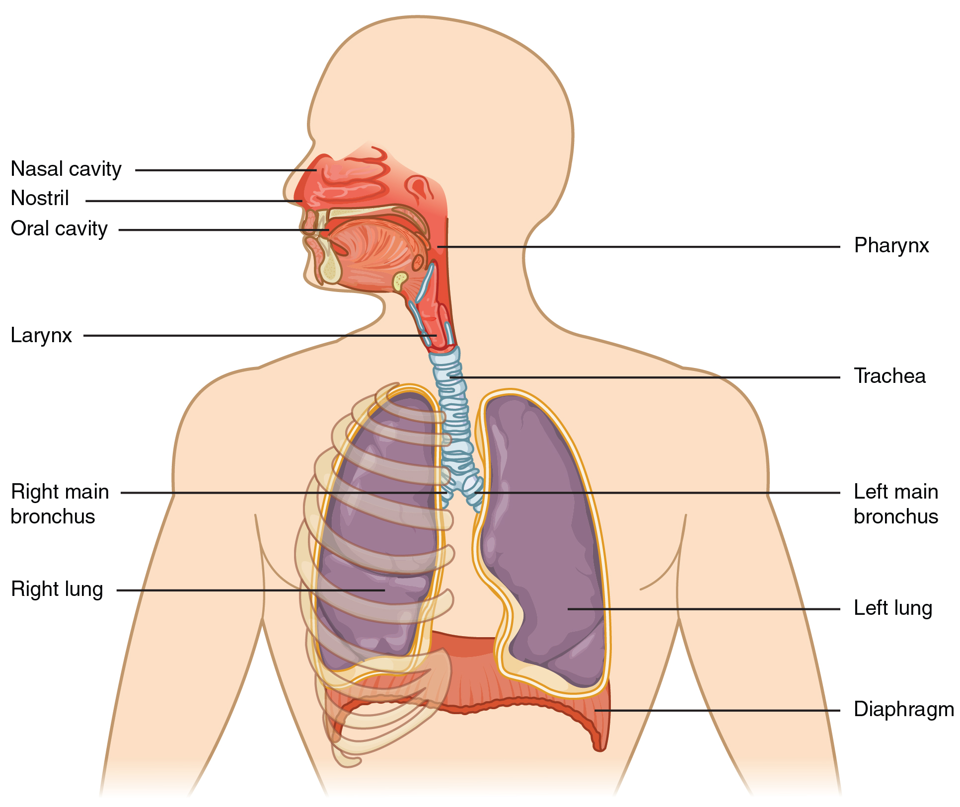
Published anatomical illustration: identify the larger airways, paired lungs and diaphragm. Use the enlargement for labels; this is not a clinical image.
Enlarge image for inspection
Scroll across the whole source image to inspect its labels. Try identification before opening the key.
Check the identification key
lungs: The two large purple organs on either side of the central airway; patient's right is image left. diaphragm: Red domed sheet beneath both lungs, indicated by the lower-right label. larynx: The labelled red and blue cartilaginous region above the ringed trachea. trachea: Vertical pale-blue ringed airway down the centre of the neck and chest. bronchi: Two branches at the bottom of the trachea entering the lungs, identified by Right main bronchus and Left main bronchus labels. ribs: Translucent curved beige bones drawn across the right lung on image left. Use the thorax figure for an explicit Ribs label. No bronchioles, alveoli, capillaries or intercostal labels. Ribs are drawn but not labelled. Anatomical left/right is the person's left/right, opposite the viewer.
Read the anatomical illustration
The published Major Respiratory Organs illustration shows a labelled larynx above the trachea, which divides into right and left main bronchi. The two lungs occupy the chest and the diaphragm is the curved muscle below them. Follow the source labels and the connected structures; the patient’s right appears on the viewer’s left in this front view.
This is an anatomical illustration rather than a photograph or a clinical scan. Purple distinguishes the lungs from the surrounding structures in this artist’s scheme. The ringed central airway helps locate the trachea, but colour and drawing size are conventions, not evidence that real lungs are purple or that this page gives measured organ dimensions.
Match the image to the route
Locate the larynx, then trace the trachea down to its division into bronchi. Find the paired lungs around these branches and the dome-shaped diaphragm below. Compare this with the original airway and chest diagrams; the relationships agree even though the illustrations use different styles and overlap more anatomy.
The image does not resolve microscopic alveoli or capillaries and does not provide labelled intercostal layers. Use the next source images for those structures instead of inventing details from this view. Try the identification questions before opening the key; enlargement retains the complete source image and makes its small labels readable.
Pause and explain
Which structure lies below the lungs in this source image?
Worked example
Locate the central airway above its two main branches.
Show the worked solution
- Follow the larynx downwards.
- Stop above the right and left main bronchi.
Answer The trachea.
Try a topic test
Choose Easy, Normal or Difficult. Save your answers and open worked review after finishing.
Test this topic →From guided practice to a new situation
Write your answer and reasoning first. Use a hint only if you are stuck. The model solution is for self-checking; your written work is not automatically marked.
Identify the paired purple organs and the curved muscle below them in the published image.
Give me a hint
Follow the lung and diaphragm source labels.
Compare my reasoning
- The paired organs are right and left lungs.
- The curved muscle beneath is the diaphragm.
Lungs; diaphragm.
Look for these in your work
- Identify both
- Use the source relationships
Name the airway above the trachea and the two main branches below it.
Give me a hint
Trace the connected route.
Compare my reasoning
- The upper structure is the larynx.
- The lower division forms right and left main bronchi.
Larynx; right and left main bronchi.
Look for these in your work
- Name all structures
- Preserve the route
Can this whole-organ image identify an individual capillary wrapped around an alveolus? Explain which source view you need.
Give me a hint
Check the level of detail.
Compare my reasoning
- The whole-chest illustration lacks the microscopic exchange network.
- The Respiratory Zone image shows the alveolus and associated capillaries.
No; use the magnified Respiratory Zone image for alveoli and associated capillaries.
Look for these in your work
- State the resolution limit
- Select the appropriate image
Common mistakes
- Mistaking viewer left for patient left.
- Claiming alveoli are resolved in a whole-chest image.
What can you explain now?
Which structure lies below the lungs in this source image?
Compare with the explanation
Diaphragm
The curved muscular boundary is labelled.
After trying it yourself, choose your next review. This is your self-assessment.
Your review choice appears on Today. Sign in to sync it across devices.
Make it stick
Locate five required organ types before checking the source key.
If you needed the solution, close it and solve the problem again from a blank page. A correct answer is stronger when you can explain the reason for each step.