Recognise asexual examples in published photographs
Identify runner and budding structures while separating observation from genetic inference.
Core and Extended; Biology 0610, 2026–2028. Use the stated specimen features and evidence. Diagrams are schematic. Written practice is self-checked
Before you begin
- Recall cells, DNA, inherited characteristics and the distinction between growth of an organism and production of offspring.
Work on paper. Try each question before revealing help, and explain why your method works.
Quick readiness check
What is directly visible in the runner photograph?
What you will learn
- Identify visible structures with the image key
- Keep static-image limits explicit
Recognise asexual examples in published photographs
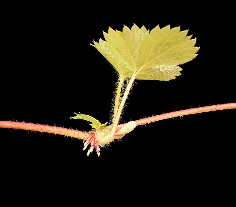
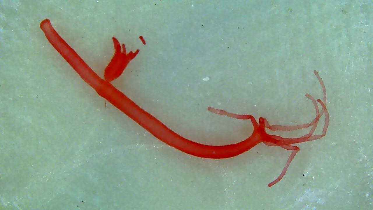
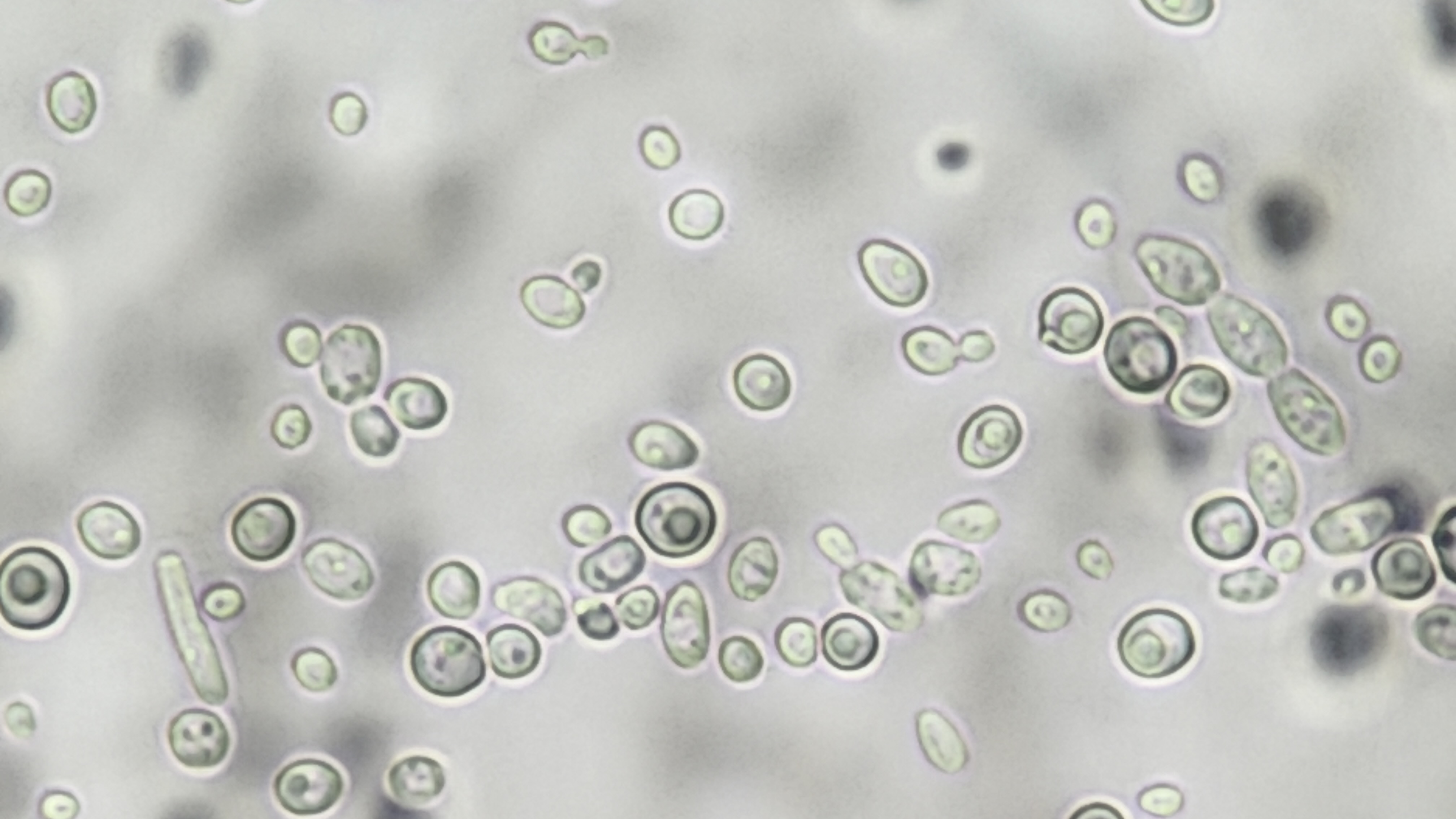
Unchanged photographs show a runner plantlet and budding hydra and yeast. Morphology supports identification; genotype, complete development and unprovided scale remain separate. Original teaching model.
Runner plantlet · Pink Panda strawberry hybrid

Enlarge image for inspection
Inspect the unchanged original at its native pixel size. Scroll within the image to follow the visible structures.
Check the identification key
- runner: Horizontal pink/red stem crossing the frame.
- daughter plantlet: Leafy shoot arising from the central node.
- developing roots: Short pale/pink projections below the same node.
Source identifies an ornamental strawberry × Comarum hybrid, Pink Panda; do not label it as pure garden strawberry or a specified strawberry species. Shows a developing plantlet with emerging roots, not an established daughter plant rooted in soil. The parent plant is outside the frame. No printed labels, scale bar, flower, seed, mitotic chromosomes or genotype are shown. 800-pixel width is suitable for normal display; enlarged root details will soften.
Hydra with an attached bud

Enlarge image for inspection
Inspect the unchanged original at its native pixel size. Scroll within the image to follow the visible structures.
Check the identification key
- parent: Larger curved red tube with long tentacles at right.
- attached bud: Smaller projection attached near upper-left centre.
- bud tentacles: Short projections at the free end of the bud.
Static image shows attachment and different sizes, not growth over time or bud detachment. Source gives genus Hydra without species identification. Magnification, scale and specimen preparation are not specified; do not infer that this is a living specimen or assign body dimensions. No nuclei, mitotic stages, fusion, DNA or genotype are identifiable.
Yeast cells with budding shapes

Enlarge image for inspection
Inspect the unchanged original at its native pixel size. Scroll within the image to follow the visible structures.
Check the identification key
- larger cell: Large oval part of an isolated unequal attached pair.
- bud: Small rounded outgrowth connected to the larger cell through a narrow neck; use the clear top-centre or centre-right examples.
- avoid: Do not identify two overlapping independent cells as a budding pair.
Source identifies yeast but does not specify species; do not call it Saccharomyces cerevisiae or Candida. The file description provides no exact magnification or scale bar; no cell-size calculations are supported. Only a static budding shape is shown, not a time sequence or detachment. Internal contrast does not reliably identify nuclei or chromosomes; do not claim mitotic stages. Image cannot establish genotype or genetic identity.
| Feature | Meaning |
|---|---|
| Runner | Pink Panda strawberry hybrid; emerging roots, parent outside frame |
| Hydra | Attached smaller bud with developing tentacles |
| Yeast | Unequal attached cell–bud pairs; species and scale unspecified |
| Limit | No photo directly establishes genotype |
Identify visible structures with the image key
The first credited photograph shows the ornamental strawberry × Comarum hybrid Pink Panda. A horizontal pink-red runner crosses the frame; a leafy daughter shoot develops from a central node, with short emerging roots below it. The parent is outside the frame, and the roots are not shown established in soil. Identify the runner, daughter shoot and emerging roots rather than describing an unseen fully rooted parent–daughter pair.
The hydra photograph shows a larger curved tubular parent with long tentacles towards the right. A smaller attached bud projects from its side near the upper-left centre, with shorter developing tentacles at the bud’s free end. This attached outgrowth supports recognition of budding. A static image does not show the entire growth sequence or later detachment of the bud.
Keep static-image limits explicit
The yeast wet-mount photograph includes larger oval cells with smaller attached rounded buds, with clear unequal pairs near the upper centre and centre-right. Many cells overlap or lie close together; do not count every neighbouring pair as a bud. Use a narrow connection and unequal outgrowth shape with the published yeast context. The source supplies no species identification, scale bar or exact magnification.
All three photographs show morphology rather than genetic information. They cannot directly prove identical genotype, absence of gamete fusion throughout a life cycle or a universal reproduction rate. Use the supplied copying history for the expected clone relationship and the image to identify visible structures. Enlargement and identification keys support observation without adding labels to or changing the originals.
Pause and explain
Which statement fits the yeast photograph?
Worked example
Identify the horizontal structure and the projections below the central leafy node in the runner photograph.
Show the worked solution
- Trace the pink-red horizontal connection.
- Distinguish the short roots emerging below the node.
Answer Runner stem and emerging roots below the daughter node; roots are not shown established in soil.
Try a topic test
Choose Easy, Normal or Difficult. Save your answers and open worked review after finishing.
Test this topic →From guided practice to a new situation
Write your answer and reasoning first. Use a hint only if you are stuck. The model solution is for self-checking; your written work is not automatically marked.
Identify three visible structures in the runner photograph.
Give me a hint
Follow the horizontal stem, leafy node and lower projections.
Compare my reasoning
- The runner is the horizontal stem.
- The node carries a leafy daughter shoot and emerging roots.
Runner stem, daughter leafy shoot at the node, and short emerging roots below it.
Look for these in your work
- Identify all three
- Do not claim an unseen parent
Locate the parent and bud in the hydra image and give a supporting feature.
Give me a hint
Compare the larger tube with the attached smaller outgrowth.
Compare my reasoning
- The larger tube has long tentacles at the right.
- The smaller attached bud has developing tentacles.
Parent: larger curved tube with long right-hand tentacles. Bud: smaller attached side outgrowth with developing tentacles.
Look for these in your work
- Locate both structures
- Use visible attachment and tentacles
Why does the yeast image support budding identification but not exact size or genotype?
Give me a hint
Separate visible outlines from missing scale and genetic information.
Compare my reasoning
- Unequal attached cells with a narrow connection support budding.
- No scale or genetic record is supplied by the photograph.
Unequal attached cell–bud shapes support identification, but the photograph lacks a scale for exact size and does not reveal genotype. The yeast species is also unspecified.
Look for these in your work
- Use morphology
- State scale and genetic limits
Common mistakes
- Claiming the runner parent is visible.
- Inferring genotype or exact size without genetic or scale evidence.
What can you explain now?
Which statement fits the yeast photograph?
Compare with the explanation
Budding shapes are visible, but species and exact size are not supplied
The source lacks species and scale information.
After trying it yourself, choose your next review. This is your self-assessment.
Your review choice appears on Today. Sign in to sync it across devices.
Make it stick
Identify a visible reproductive structure in each photograph and state one conclusion the image cannot support.
If you needed the solution, close it and solve the problem again from a blank page. A correct answer is stronger when you can explain the reason for each step.