Identify ribs and intercostal muscles in a published image
Recognise bone and between-rib muscle in the source thorax illustration.
Core and Extended; Biology 0610, 2026–2028. Use the stated specimen features and evidence. Diagrams are schematic. Written practice is self-checked
Before you begin
- Recall the location of intercostal muscles.
Work on paper. Try each question before revealing help, and explain why your method works.
Quick readiness check
Where do intercostal muscles lie?
What you will learn
- Read bone and muscle landmarks
- Connect views without inventing motion
Identify ribs and intercostal muscles in a published image
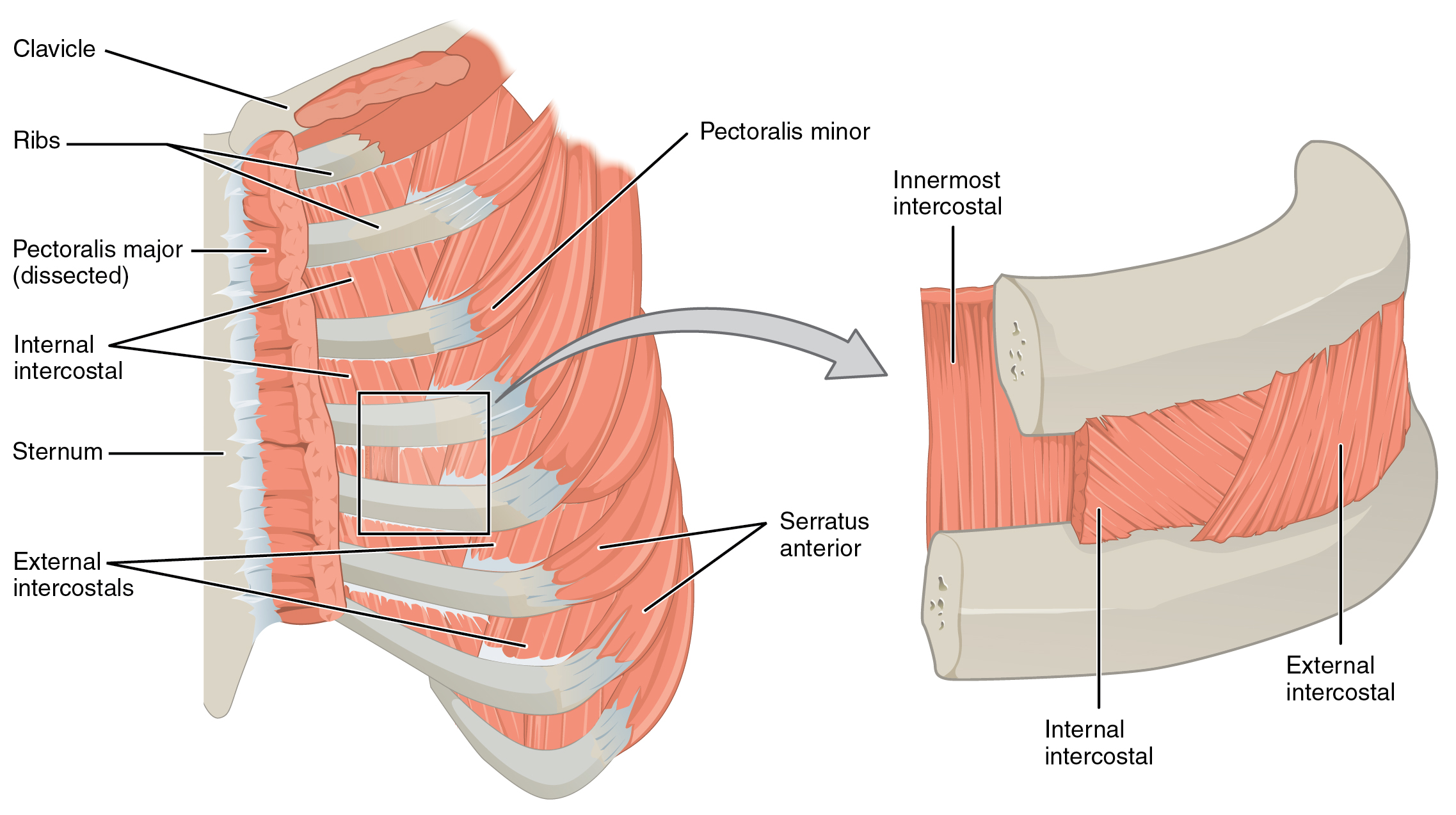
The source thorax illustration separates pale rib bones from intercostal muscles between them; it does not show the lungs or diaphragm.
Enlarge image for inspection
Scroll across the whole source image to inspect its labels. Try identification before opening the key.
Check the identification key
ribs: Pale curved bones labelled Ribs on image left; magnified upper and lower bone sections on image right. internal intercostals: Muscle between ribs near the sternum in the main panel; labelled middle layer with diagonal fibres in the magnified panel. external intercostals: Muscle between ribs farther from the sternum in the main panel; labelled outermost layer on the right of the magnified panel, with fibres crossing the internal layer's direction. Includes the additional Innermost intercostal label, which is beyond the requested syllabus. The layer view demonstrates position and fibre direction, not contraction or rib movement. No diaphragm or lungs shown.
Read bone and muscle landmarks
The published Thorax illustration shows pale curved ribs and muscles spanning the spaces between them. The main panel and its magnified layer view provide different levels of detail. For Core identification, recognise ribs as bones and intercostal muscles as the tissue between ribs; do not label an entire rib as muscle.
The main panel includes the sternum towards the front and shows muscle regions at different positions. These additional landmarks help orientation, but the required answer here remains ribs and intercostal muscles. A tissue band between adjacent bones is located using its endpoints and the source label rather than a memorised page coordinate.
Connect views without inventing motion
The enlargement shows several muscle layers between the same pair of ribs. Internal and external layers are distinguished in a later Extended lesson. The original illustration also labels an innermost layer; this extra anatomical detail does not change the Core distinction between rib bone and between-rib muscle.
The image is a labelled anatomical illustration, not a moving record of a breath. It establishes positions and layers but does not by itself show which muscle is contracting or whether a person is inspiring. Lungs and the diaphragm are absent from this isolated thorax-muscle view; use the Major Respiratory Organs image for them.
Pause and explain
What does this still source image directly establish?
Worked example
Identify the pale curved structures and the bands between them.
Show the worked solution
- Read the Ribs label.
- The between-rib tissue is muscle.
Answer Ribs; intercostal muscles.
Try a topic test
Choose Easy, Normal or Difficult. Save your answers and open worked review after finishing.
Test this topic →From guided practice to a new situation
Write your answer and reasoning first. Use a hint only if you are stuck. The model solution is for self-checking; your written work is not automatically marked.
Identify ribs and intercostal muscles in the published Thorax image.
Give me a hint
Use bone and between-bone tissue.
Compare my reasoning
- Pale curved bones are ribs.
- Tissue spanning the spaces is intercostal muscle.
Ribs are the pale curved bones; intercostal muscles occupy the spaces between them.
Look for these in your work
- Identify both
- Use the anatomical relationship
What does the magnified panel add to the main panel?
Give me a hint
Compare its level of detail.
Compare my reasoning
- The main panel places muscles in the chest wall.
- The magnification separates between-rib muscle layers.
It shows muscle layers between adjacent ribs more clearly.
Look for these in your work
- Compare the views
- Avoid inventing motion
A student claims the diaphragm is visible because every respiratory image contains it. Evaluate the claim for this source.
Give me a hint
Use this image’s actual content.
Compare my reasoning
- This isolated muscle view shows ribs and intercostals.
- It does not show the diaphragm.
The claim is unsupported; use the separate Major Respiratory Organs image to identify the diaphragm.
Look for these in your work
- State the actual scope
- Select another view
Common mistakes
- Calling a rib a muscle layer.
- Inferring inspiration from a still anatomy image.
What can you explain now?
What does this still source image directly establish?
Compare with the explanation
Positions of ribs and intercostal muscle layers
Its labels and landmarks show anatomy.
After trying it yourself, choose your next review. This is your self-assessment.
Your review choice appears on Today. Sign in to sync it across devices.
Make it stick
Identify bone and muscle in both panels without inferring a breathing phase.
If you needed the solution, close it and solve the problem again from a blank page. A correct answer is stronger when you can explain the reason for each step.